A neuroscientist conducted a pioneering study by scanning her own brain 26 times before, during, and after pregnancy to observe physical changes, revealing decreases in gray matter and improvements in white matter, though the implications of these changes remain unclear. The research aims to better understand brain adaptations during pregnancy and their potential links to postpartum depression.
A study from the University of Wurzburg reveals that intelligence is a global property of the brain, influenced by distributed connections rather than specific regions like the prefrontal cortex. Using fMRI data from over 800 individuals, researchers found that brain-wide connectivity better predicts general, crystallized, and fluid intelligence. This challenges existing models focused on localized brain areas and suggests that intelligence involves complex, system-wide brain interactions.
Researchers have discovered that the brain's default mode network (DMN) initiates creative ideas, which are then evaluated by other brain regions. Using advanced brain imaging, they tracked real-time brain activity during creative tasks, revealing the DMN's crucial role in creativity. This understanding could lead to interventions that enhance creativity and aid in treating mental health disorders.
A study by Ben-Gurion University reveals that people underutilize their visual working memory (VWM), often recalling fewer items than their capacity allows. Participants typically remembered just one item at a time, despite being able to remember 3-4 items. This finding provides new insights into the real-life usage of VWM and suggests that further research is needed to understand the reasons behind this behavior.
A neuroimaging study in South Korea found that individuals with social anxiety disorder have increased cortical thickness in certain brain regions, along with reduced neuron numbers in areas related to attention and socio-emotional processing. The study, published in Psychiatry Research: Neuroimaging, identified alterations in the insula, superior parietal lobule, frontopolar cortex, and superior temporal gyrus, as well as reduced thickness in the left superior/middle frontal gyrus and left fusiform gyrus. These findings suggest distinct neural mechanisms underlying social anxiety disorder, although further research is needed to confirm the results.
Researchers at the University of Colorado have identified three types of drug inhibitors that can regulate the CaMKII protein, which plays a crucial role in brain and heart function, to potentially mitigate Alzheimer's and heart conditions. These inhibitors offer a valuable tool for scientists to study the protein's functions and could lead to significant progress in treating brain diseases without harmful side effects. The research provides accessible tools for scientists across various disciplines to explore the protein's roles and functions.
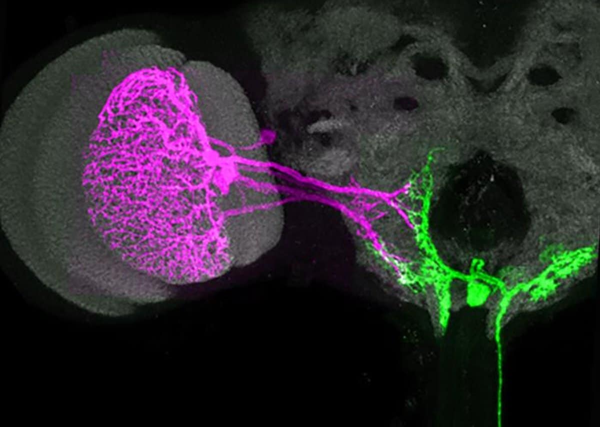
A groundbreaking study on fruit flies has challenged traditional views on motor neurons, revealing that individual motor neurons can produce a variety of complex head movements rather than just simple actions. Utilizing advanced laboratory techniques and artificial intelligence, researchers were able to stimulate single motor neurons and observe the resultant movements, uncovering a sophisticated system akin to a digital thermostat that adjusts based on the body’s current posture. This discovery not only challenges existing notions of motor neuron functionality but also opens new avenues for understanding motor system diseases and the interplay between different types of neurons in movement control.
A study explores how the brain perceives cooling sensations, particularly when consuming cold foods like mint cookies, by focusing on TRPM8 receptors in the mouth. Removing these receptors in mice blurs the distinction between cool and warm sensations, impacting temperature preferences. The research sheds light on temperature perception's impact on taste and dietary choices, offering insights into sensory processing and potential health implications.
A new study published in Psychiatry Research suggests that individuals using antidepressants exhibit increased dynamics in brain network connectivity, indicating that antidepressant treatment may enhance the brain’s network flexibility and integration across various regions. Contrary to initial expectations, the study found no significant differences in the dynamic reconfiguration of brain networks between individuals with major depressive disorder (MDD), anxiety disorders, or both, and those without these conditions. However, individuals using antidepressants showed even higher levels of dynamic brain network changes compared to control participants, indicating enhanced promiscuity and flexibility in connectivity patterns across the brain. The findings provide new insights into the neurobiological effects of antidepressants and highlight the importance of exploring the dynamic nature of brain connectivity in mental health disorders.
Researchers at Brown University's Carney Institute for Brain Science have uncovered the brain's ability to separately control the enhancement of relevant information and the filtering out of distractions, akin to coordinating muscles for physical tasks. Using functional magnetic resonance imaging (fMRI), they found that the anterior cingulate cortex and the intraparietal sulcus work together to adjust focus and filter settings, shedding light on human attention flexibility and potential implications for attention-related disorders like ADHD. This breakthrough offers new insights into how the brain manages to focus in noisy environments and provides a deeper understanding of attention mechanisms.
A study from Washington University reveals that during sleep, brainwaves facilitate the movement of cerebrospinal fluid through the brain, effectively flushing out waste and potentially preventing neurological diseases like Alzheimer's and Parkinson's. Understanding and enhancing this cleansing process could lead to improved sleep quality and overall brain health, offering new avenues for treating sleep disorders and combating neurodegenerative diseases.
A groundbreaking study led by researchers from the Singapore Institute for Clinical Sciences suggests that early life adversity (ELA) accelerates brain development during the critical preschool years, potentially predisposing children to adverse cognitive and mental health outcomes later in life. The study, detailed in the journal Nature Mental Health, utilized neuroimaging data from the Growing Up in Singapore Towards healthy Outcomes (GUSTO) birth cohort to track the developmental trajectory of the brain over time and found evidence of accelerated brain development among children exposed to high levels of ELA. This accelerated development was most pronounced during the preschool years and was linked to greater risk of exhibiting behavioral and emotional problems. The findings imply that while accelerated brain development in response to ELA may serve as an adaptive mechanism to cope with adverse conditions, it comes with potential costs, limiting the window for neuroplasticity and predisposing individuals to cognitive and mental health challenges later in life.
Researchers at the University of California, Irvine have developed 20 novel recombinant rabies viral vectors that offer enhanced capabilities for neural circuit mapping, particularly in aging and Alzheimer’s disease studies. These vectors can detect microstructural changes in brain neurons and target specific components of neuron biology, aiding in the analysis of pathological changes in brain diseases. The research team plans to make these innovative tools available to the neuroscience community through UCI’s Center for Neural Circuit Mapping, opening new pathways for targeted treatment strategies.
A study published in eLife reveals that older adults who perform better on cognitive tasks may be benefiting from a unique form of brain adaptability, particularly within a region involved in visual attention. The research suggests that the brain may tap into unused regions to offset age-related cognitive decline, with the cuneal cortex showing increased activity correlated with both advancing age and cognitive performance levels. This increased reliance on the cuneal cortex could reflect a strategic adaptation of the brain, potentially facilitating a more focused attention to visual aspects. However, the study's design does not allow for a clear understanding of whether the increased activation in the cuneal cortex directly causes improved cognitive performance or is simply associated with it.
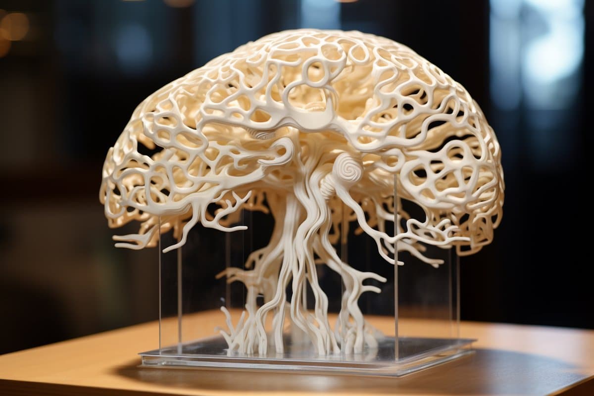
Researchers at the University of Wisconsin have developed the world's first 3D-printed brain tissue that mimics natural brain tissue, allowing neurons to interconnect and form networks similar to human brain structures. This breakthrough technique offers unparalleled opportunities to study brain functions and disorders, impacting the understanding and treatment of neurological conditions such as Alzheimer's and Parkinson's. The 3D-printed brain tissue can form networks and communicate through neurotransmitters, providing a powerful model for studying brain cells and communication in humans.