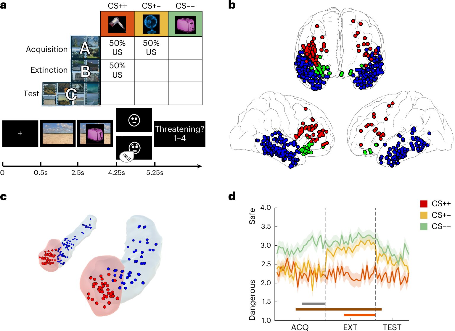
A study published in Nature Human Behavior reveals that fear extinction involves stable, context-specific neural representations in brain regions like the amygdala and hippocampus, with implications for understanding and treating anxiety disorders and phobias.
The brain seamlessly switches between external perception and internal memory, involving overlapping yet distinct neural networks, with ongoing research exploring whether a central control system manages these shifts or if they emerge from competitive dynamics within shared networks, which has implications for understanding cognitive function and disorders.
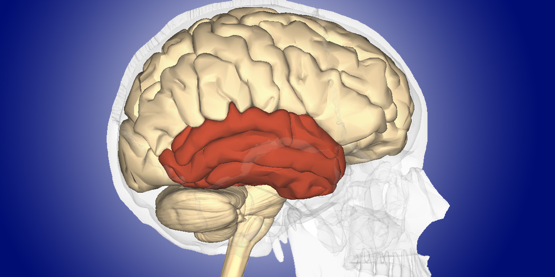
A study in Neuropsychologia reveals that adults who stutter show lower accuracy in repeating nonword syllable sequences, linked to distinct brain activity patterns in sensorimotor and auditory regions. This research provides insights into the neural mechanisms of stuttering, potentially aiding in the development of better therapeutic strategies.
A neuroimaging study in South Korea found that individuals with social anxiety disorder have increased cortical thickness in certain brain regions, along with reduced neuron numbers in areas related to attention and socio-emotional processing. The study, published in Psychiatry Research: Neuroimaging, identified alterations in the insula, superior parietal lobule, frontopolar cortex, and superior temporal gyrus, as well as reduced thickness in the left superior/middle frontal gyrus and left fusiform gyrus. These findings suggest distinct neural mechanisms underlying social anxiety disorder, although further research is needed to confirm the results.
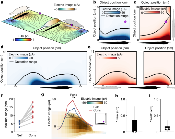
Research on electric fish reveals insights into collective sensing and communication in active-sensing animals, shedding light on the neural mechanisms and evolution of behavior. The study explores distributed information fusion in multistatic sensor networks for underwater surveillance and the potential for group hunting via eavesdropping in echolocating bats, as well as the evidence for mutual allocation of social attention through interactive signaling in weakly electric fish. This research contributes to a better understanding of how animals use their sensory systems to navigate and communicate in complex environments.
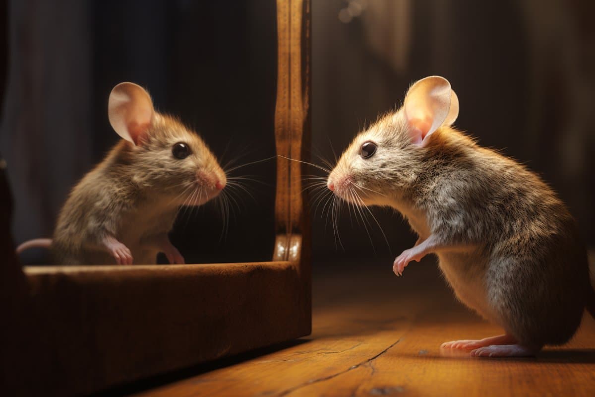
Mice exhibit behavior resembling self-recognition when viewing themselves in mirrors, but only under specific conditions such as familiarity with mirrors, socialization with similar-looking mice, and visible markings on their fur. Researchers have identified a subset of neurons in the hippocampus that are crucial for this self-recognition-like behavior. The study highlights the importance of social experiences and sensory cues in developing self-recognition capabilities, providing valuable insights into the neural mechanisms behind self-recognition.
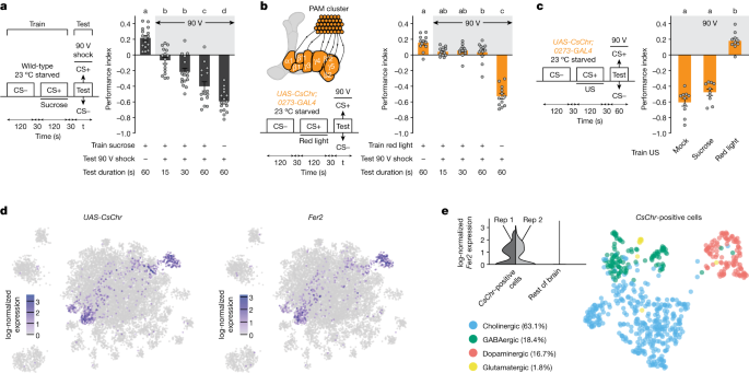
Researchers have discovered that specific dopaminergic neurons in the brains of fruit flies play a crucial role in driving reward-seeking behavior, even in the face of adverse consequences. The study found that activation of these neurons, known as β′2 and γ4 DANs, led the flies to persistently seek rewards, such as sucrose, despite being subjected to electric shocks. The findings shed light on the neural mechanisms underlying reward-seeking behavior and could have implications for understanding substance use disorders in humans.
A study published in Biological Psychiatry: Cognitive Neuroscience and Neuroimaging has revealed differences in functional brain connectivity in individuals with schizophrenia, shedding light on the neural basis of the disorder. Using advanced brain imaging and mathematical techniques, researchers found impairment in the organizational pattern that differentiates visual and sensorimotor pathways in those with schizophrenia. This impairment was linked to the clinical symptoms of the disease, providing insights into its mechanisms. The findings suggest that changes in brain organization may offer valuable insights into the progression and mechanisms of schizophrenia, potentially leading to new approaches for treatment.
A new neuroimaging study provides evidence of increased amygdala and face cortical network activation in individuals with autism spectrum disorder (ASD) in response to face-like (or pareidolic) stimuli. These findings support the hypothesis of an overly connected subcortical face-processing network in ASD, potentially resulting from an early imbalance between excitatory and inhibitory systems. The researchers propose that the increased sensitivity of the amygdala in ASD for pareidolic objects may be evidence of over-connection between the amygdala and the rest of the face-processing system. This over-connection could result from an early imbalance between excitatory and inhibitory systems in autism.
Researchers used intracranial recordings to study the neural mechanisms underlying humor processing and found that high-frequency brain waves increased during the funniest scenes in a Charlie Chaplin clip. The study revealed consistent involvement of the anterior temporal lobe, the temporo-parietal junction, and the temporal-occipital sulcus in humor processing. The findings support the idea that humor processing involves two complementary mechanisms: the detection of incongruity and the experience of positive emotions. The study provides insights into the neural correlates of humor processing and highlights the role of the temporal lobe in humor.