Tag

Brain Imaging

All articles tagged with #brain imaging

Brain Changes Tie Hearing Loss to Dementia Risk, Study Finds
health9 days ago

Brain Changes Tie Hearing Loss to Dementia Risk, Study Finds

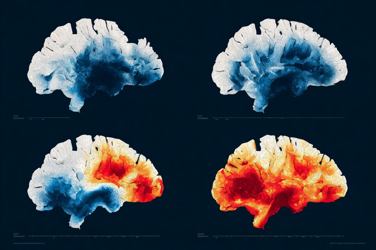
A new study links age-related hearing loss (presbycusis) to cognitive decline through coupled functional and structural brain changes. Researchers introduce the Functional-Structural Ratio (FSR), derived from MRI measures, which correlates with worse hearing and poorer cognitive performance in specific brain regions, suggesting FSR could serve as a biomarker for dementia risk. While cross-sectional, the findings highlight the potential of preserving hearing health to protect brain integrity and guide early interventions.

Moderate cannabis use linked to larger brain regions and better cognition in middle-aged and older adults
health22 days ago

Moderate cannabis use linked to larger brain regions and better cognition in middle-aged and older adults

A UK Biobank analysis of 26,362 adults aged 40–77 finds that greater lifetime cannabis use is generally associated with larger regional brain volumes and better cognitive performance, especially with moderate use; some measures peak at high use, but one region (posterior cingulate) shows lower volume with more use, and sex differences appear but are not consistent. The study emphasizes nuance, cautions against causal conclusions, and notes context (patterns, products, timing) matters, with future work on brain connectivity and other substances like psilocybin.

When the mind’s eye goes dim: what aphantasia reveals about imagination
science24 days ago

When the mind’s eye goes dim: what aphantasia reveals about imagination

Most people can visualize details from daily life, but about 4% have little to no mental imagery (aphantasia). The article traces its discovery, how researchers measure imagery (e.g., binocular rivalry) and brain activity, and suggests aphantasia may involve both lack of conscious access and difficulty in voluntary generation. Despite variability and possible genetic links, many with aphantasia function normally in memory and creativity, making it a valuable window into how the brain constructs inner experiences and consciousness.

New 3D Color Imaging Joins Ultrasound and Photoacoustics to See Inside the Body
technology1 month ago

New 3D Color Imaging Joins Ultrasound and Photoacoustics to See Inside the Body

Caltech and USC researchers have developed RUS-PAT, a hybrid rotational ultrasound and photoacoustic tomography system that produces fast, three-dimensional color images showing both tissue structure and blood-vessel function. Demonstrations across multiple body regions suggest wide medical potential, including enhanced breast tumor imaging, monitoring nerve damage from diabetes, and concurrent brain structure and blood flow visualization. The technique reaches about 4 cm depth and can complete a scan in under a minute, with ongoing translational development for clinical use.

Lasting brain changes found in recovered COVID-19 patients, MRI study shows
neuroscience1 month ago

Lasting brain changes found in recovered COVID-19 patients, MRI study shows

A multimodal MRI study comparing Long COVID, recovered (symptom-free), and never-infected groups (47 participants) found distinct brain changes in tissue microstructure and neurochemistry associated with Long COVID and footprints in those who have fully recovered. Differences include altered myelin proxies (T1w/T2w) in motor and memory regions, diffusion changes in the cerebellum and brainstem, and metabolic shifts (lower glutamine, higher N-acetyl-aspartate) that correlated with physical function and cognitive scores. The study suggests SARS-CoV-2 may leave a brain footprint even after apparent recovery, but its cross-sectional design and small sample size mean longitudinal research is needed to determine permanence and timeline.

Sudoku Triggers Reflex Seizures in Hypoxia-Linked Brain Case
health1 month ago

Sudoku Triggers Reflex Seizures in Hypoxia-Linked Brain Case

After surviving an avalanche that caused hypoxia, a 25-year-old German man later developed rhythmic jerks in his left arm specifically while solving Sudoku. EEG showed a right centroparietal seizure pattern; MRI was normal, but functional MRI revealed widespread brain activation with fewer inhibitory fibers in the centroparietal region, linking the hypoxia‑induced brain damage to reflex seizures triggered by visual‑spatial tasks. He was treated with anti‑epileptic medication and rehab, and remained seizure‑free for years, even giving up solving Sudoku.

Widespread brain shrinkage drives aging-related memory decline, mega-analysis finds
health-and-medicine1 month ago

Widespread brain shrinkage drives aging-related memory decline, mega-analysis finds

An international mega-analysis of over 10,000 MRI scans and 13,000 memory tests from thousands of healthy adults shows that memory decline with age stems from broad structural brain changes across many regions—especially, but not exclusively, the hippocampus—with nonlinear acceleration in later life and not fully explained by Alzheimer's risk genes like APOE ε4. This points to a distributed brain vulnerability that could help identify at-risk individuals and guide personalized cognitive health interventions.

New Brain Research Changes Understanding of How ADHD Medications Work
health2 months ago

New Brain Research Changes Understanding of How ADHD Medications Work

A large brain imaging study reveals that ADHD medications like Adderall and Ritalin primarily affect brain regions related to arousal and wakefulness, not attention networks as previously believed. These drugs mimic the brain patterns of good sleep, effectively rescuing sleep-deprived children temporarily, but do not enhance cognitive abilities in well-rested children. The findings suggest that sleep issues may underlie some ADHD symptoms and highlight the importance of addressing sleep health.

Challenges in Interpreting BOLD MRI Signals and Brain Metabolism
science2 months ago

Challenges in Interpreting BOLD MRI Signals and Brain Metabolism

Recent research reveals that fMRI signals often misrepresent actual neural activity, with about 40% of cases showing increased signals where neural activity is reduced, due to regions extracting more oxygen without increased blood flow. This challenges long-standing assumptions in brain imaging and suggests a need for direct energy consumption measurements to better understand brain function and disorders.

Couples Experience Shared Memory Loss, Study Finds
science2 months ago

Couples Experience Shared Memory Loss, Study Finds

New research shows that romantic partners synchronize their brain activity during storytelling, leading to a shared form of forgetting where they tend to forget related details, a phenomenon less observed among strangers. This neural alignment, especially in the prefrontal cortex, facilitates a shared reality but may cause loss of individual memories, highlighting how close relationships influence cognitive processes.