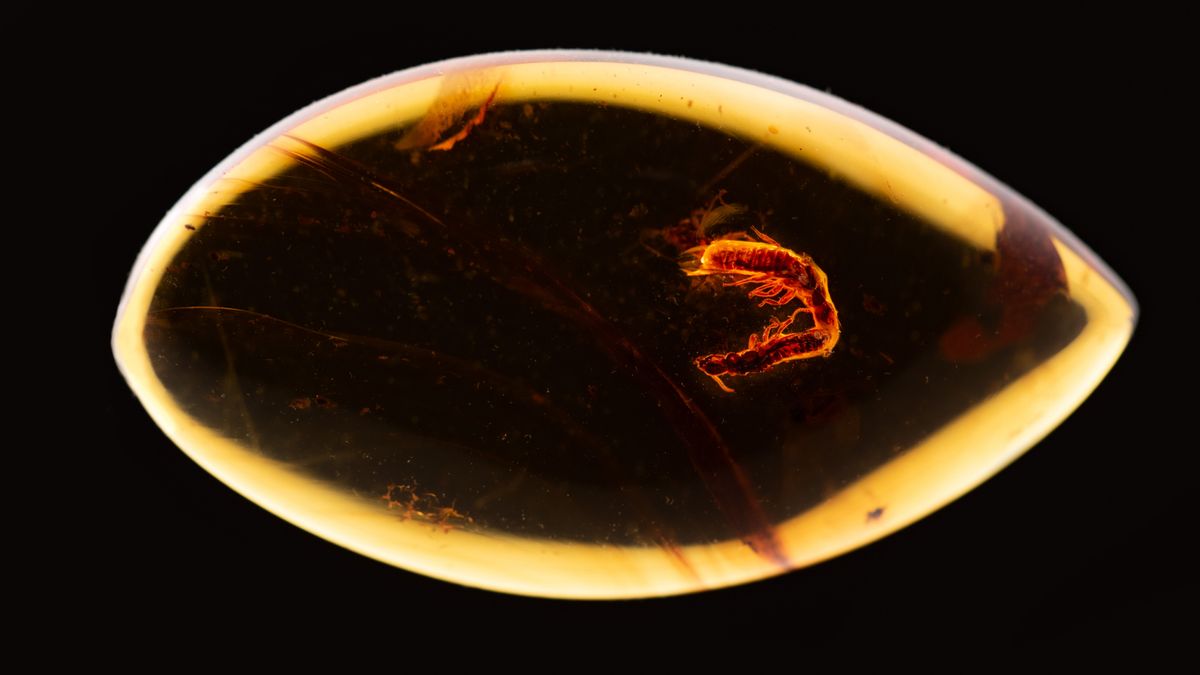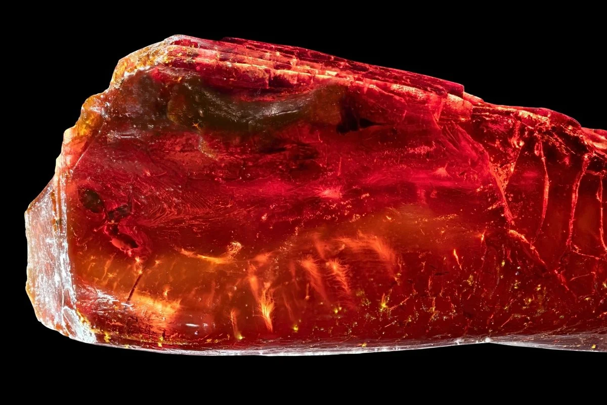
Ultra-detailed ancient ant revealed in amber through 3D imaging
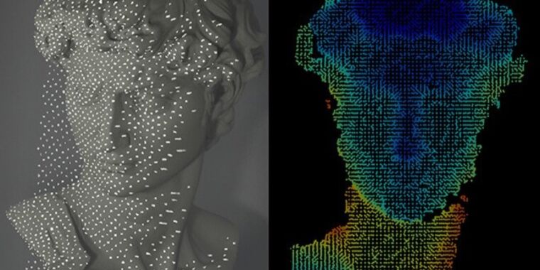
Researchers studied two amber pieces from the Goethe National Museum containing a fungus gnat, a black fly, and an extinct ant, †Ctenobethylus goepperti. Using advanced 3D imaging, they reconstructed the ant’s morphology and interior anatomy, revealing fine body hairs and enabling detailed comparisons with living Liometopum ants to infer possible canopy-nesting behavior. The findings, published in Scientific Reports, provide a clearer picture of how this ancient species lived.