A Nature Neuroscience study reveals a mathematical flaw in lesion network mapping, producing nearly identical brain networks across diverse conditions and challenging the validity of disease-specific targets, prompting calls for revised methods and cautious clinical application.
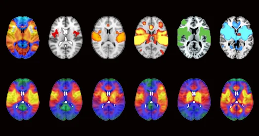
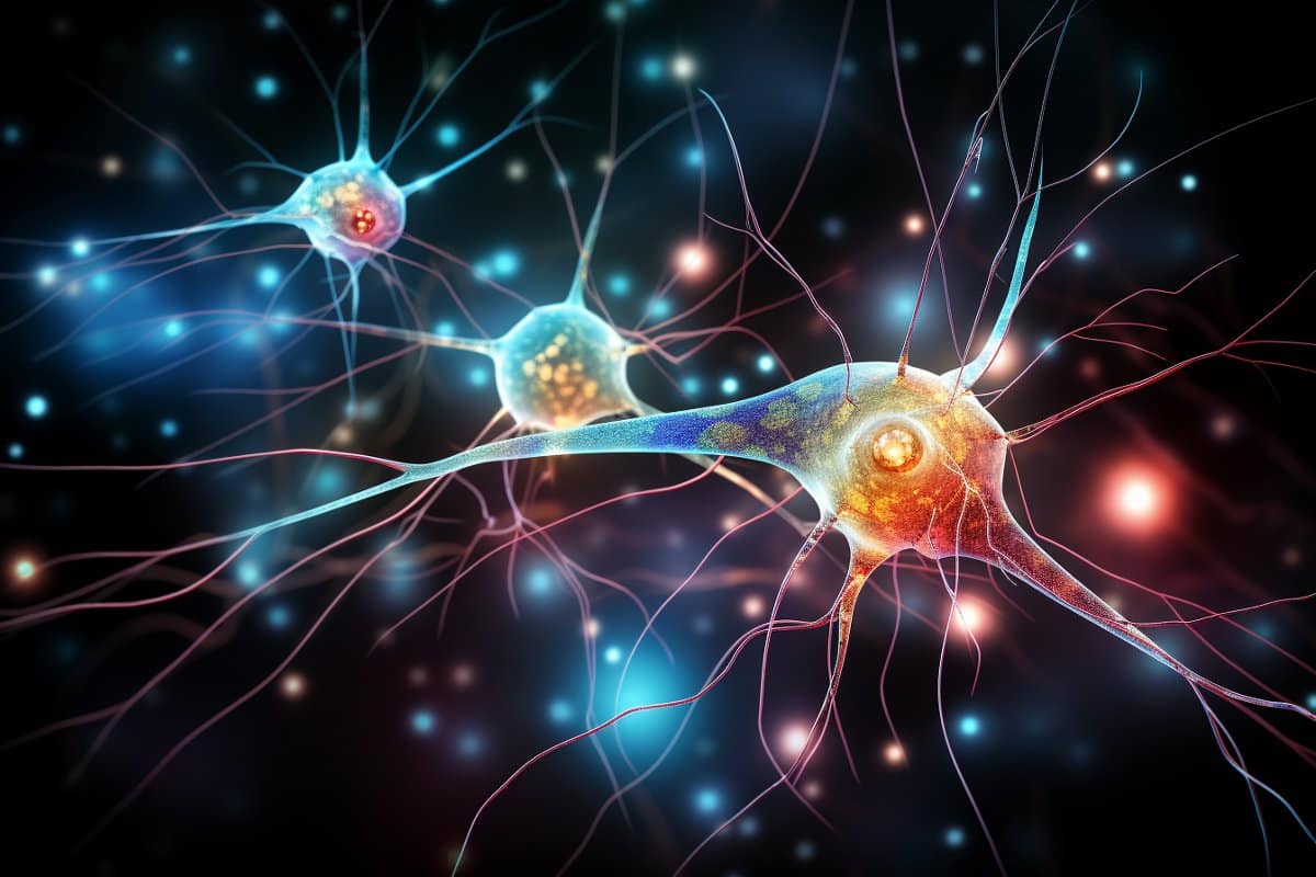
New research using MRI data and meta-analysis tools reveals that the brain's connectivity patterns serve as unique fingerprints for different regions, strongly linking structure to function across various mental activities, providing a comprehensive baseline for understanding healthy brain organization and potential neurological disorders.
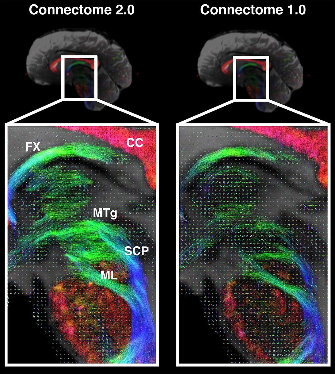
Scientists supported by NIH have developed the Connectome 2.0, an ultra-high-resolution MRI scanner capable of noninvasively imaging microscopic brain structures, advancing our ability to map the brain's wiring and understand neurological disorders at a cellular level.
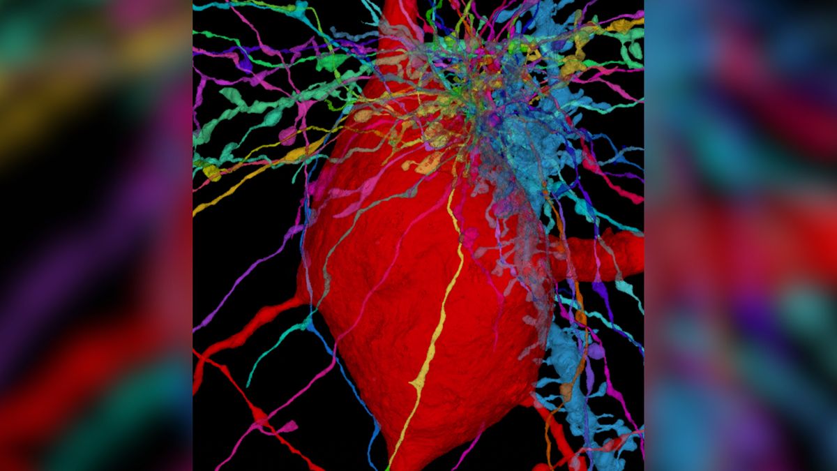
Scientists have created a detailed 3D map of the primary cilia, tiny hairlike structures found on the surface of brain cells, across the human cortex. These cilia act as antennae, sensing signals from the environment and passing them to the cell's nucleus. The map, which details 56,000 cells, could guide research into ciliopathies, diseases caused by disruptions in cilia function. The study revealed that cilia differ in size and shape depending on the cell type and cortical layer, and they are embedded in the brain's connectome. Further research will explore how primary cilia influence neural circuits and their potential use in treating neurological disorders.
Researchers at Princeton University have used the transparent worm, Caenorhabditis elegans, to study how neural information flows in the brain. By employing advanced techniques like optogenetics, they were able to visually track signal flow in real-time, neuron by neuron, and discovered unexpected "wireless signals" involving molecular releases that affect neural dynamics. This groundbreaking research challenges existing predictions based on the worm's connectome and provides valuable insights into understanding neural response.
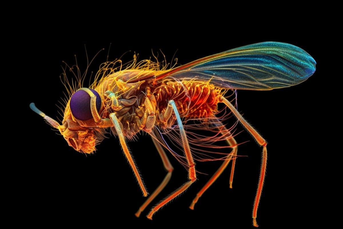
Researchers have released the most comprehensive connectome of the adult fruit fly nerve cord, providing an exceptional resource for the scientific community. The connectome, constructed from about 23,000 neurons, reveals the intricate network controlling the fly’s motor functions. New insights have already emerged from the data, challenging previous theories on fly movement. This achievement not only advances understanding of fruit fly neurology, but also serves as a model for similar future projects.