New 3D Color Imaging Joins Ultrasound and Photoacoustics to See Inside the Body
TL;DR Summary
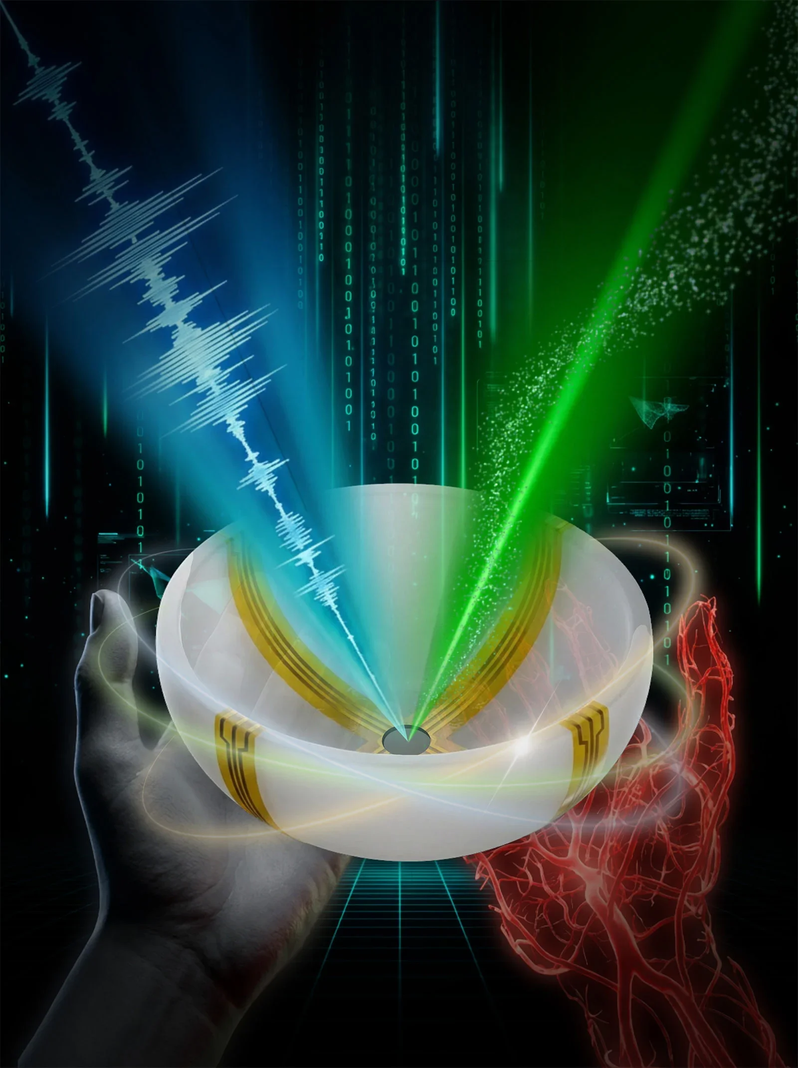
Caltech and USC researchers have developed RUS-PAT, a hybrid rotational ultrasound and photoacoustic tomography system that produces fast, three-dimensional color images showing both tissue structure and blood-vessel function. Demonstrations across multiple body regions suggest wide medical potential, including enhanced breast tumor imaging, monitoring nerve damage from diabetes, and concurrent brain structure and blood flow visualization. The technique reaches about 4 cm depth and can complete a scan in under a minute, with ongoing translational development for clinical use.
Topics:health#3d-imaging#biomedical-imaging#brain-imaging#photoacoustic-tomography#technology#ultrasound
- Scientists Develop a New Way To See Inside the Human Body Using 3D Color Imaging SciTechDaily
- Bringing Optical Color to Ultrasound Technology Org
- Optical colour ultrasound: 3D imaging technique for high-resolution diagnostics Open Access Government
- Revolutionary 3D Scanner Explores the Entire Human Body! Glass Almanac
Reading Insights
Total Reads
1
Unique Readers
15
Time Saved
9 min
vs 10 min read
Condensed
96%
1,811 → 78 words
Want the full story? Read the original article
Read on SciTechDaily