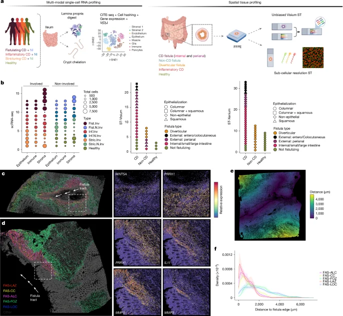
This study uses spatial transcriptomics and single-cell analysis to uncover how specialized fibroblast niches, particularly FAS cells, contribute to the formation, progression, and persistence of Crohn's fistulae, highlighting their roles in tissue remodeling, immune regulation, and epithelial regeneration, with implications for targeted therapies.
This study provides a detailed spatiotemporal map of gene expression and cellular dynamics during early human heart development, analyzing over 69,000 tissue spots and nearly 77,000 cells across 36 hearts, revealing diverse cell types, specialized conduction system components, and the formation of cardiac innervation and valves, thereby advancing understanding of human cardiogenesis.
This study used spatial transcriptomics, proteomics, and genomics to analyze the tumor immune microenvironments in diffuse large B-cell lymphoma (DLBCL), identifying distinct cellular neighborhoods and communication patterns that influence immune cell function and tumor behavior, with implications for targeted immunotherapy.
Researchers developed spVelo, a new method using neural networks to improve the measurement of gene expression changes and cell fate decisions by incorporating spatial and batch information, enabling more accurate and comprehensive analysis of cellular development and differentiation.
The article introduces spatial-DMT, a novel technology for simultaneous spatial profiling of DNA methylome and transcriptome in tissue sections at near single-cell resolution, applied to mouse embryos and brains, revealing intricate gene regulation mechanisms during development and tissue-specific epigenetic landscapes.
This study presents a comprehensive single-nucleus and spatial transcriptomic atlas of the Arabidopsis plant life cycle, revealing diverse molecular identities, cell types, and states across various organs and developmental stages, and demonstrating the power of combined technologies to uncover cellular complexity and function in plant biology.
Researchers have created a comprehensive atlas of human embryonic limb development using single-cell transcriptomic RNA sequencing and spatial transcriptomic sequencing. The study identified 67 distinct cell clusters and mapped their spatial distribution across four timepoints during the first trimester of development. The research sheds light on the cellular heterogeneity, patterning events, and gene expression patterns associated with limb malformations. The findings also highlight the similarities between human limb development and that of model organisms like mice. This atlas provides valuable insights into the complex processes involved in limb development and can serve as a reference for studying genetic variations and developmental disorders.
A study conducted by researchers at the University of Michigan has revealed new insights into the complex inflammatory response that occurs within fat tissue in obesity. Using single cell analysis and spatial transcriptomics, the study identified previously unrecognized immune cell types and interactions within adipose tissue. The researchers discovered different subtypes of macrophages, with some exhibiting pro-inflammatory genes and others showing low pro-inflammatory gene expression. The findings suggest that the body may attempt to counter inflammation by promoting lipid-associated macrophages. Further research will focus on understanding the signaling processes and proteins associated with the development of these macrophages and metabolic disorders.
Researchers are making significant progress in mapping the mouse brain by combining high-throughput single-cell RNA sequencing with spatial transcriptomics. These methods allow for the identification and mapping of different categories of brain cells, providing comprehensive atlases of the mouse brain. The next steps involve understanding the functions of these molecularly defined cell types and creating a unified resource for the neuroscience community. These efforts are part of the larger BRAIN Initiative Cell Census Network, which aims to create comprehensive maps of cells in the brains of mice and primates, including humans.
Researchers at Stanford have developed a new technique called expansion spatial transcriptomics (Ex-ST) that significantly improves the resolution of spatial transcriptomics, a method used to identify and classify different types of brain cells based on gene expression patterns. By expanding brain tissue, the researchers were able to capture mRNA from individual cells with greater precision, allowing for a more detailed understanding of neuron types and their functions. The Ex-ST method is expected to have a significant impact on neuroscience research and may pave the way for further advancements in studying the structure and function of brain tissue.
Making computational methods accessible to others is crucial for scientific progress. Jean Fan, an assistant professor at Johns Hopkins University, developed a method called STdeconvolve to analyze spatial transcriptomics data. She made the code available on GitHub and simplified installation through Bioconductor. She also shared a video of herself live-coding a spatial transcriptomics data analysis on YouTube and shared blog posts and tutorials to support students. By increasing accessibility, STdeconvolve is easier to maintain and use, and students can provide feedback and build communities.