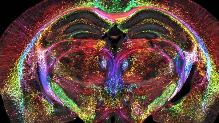
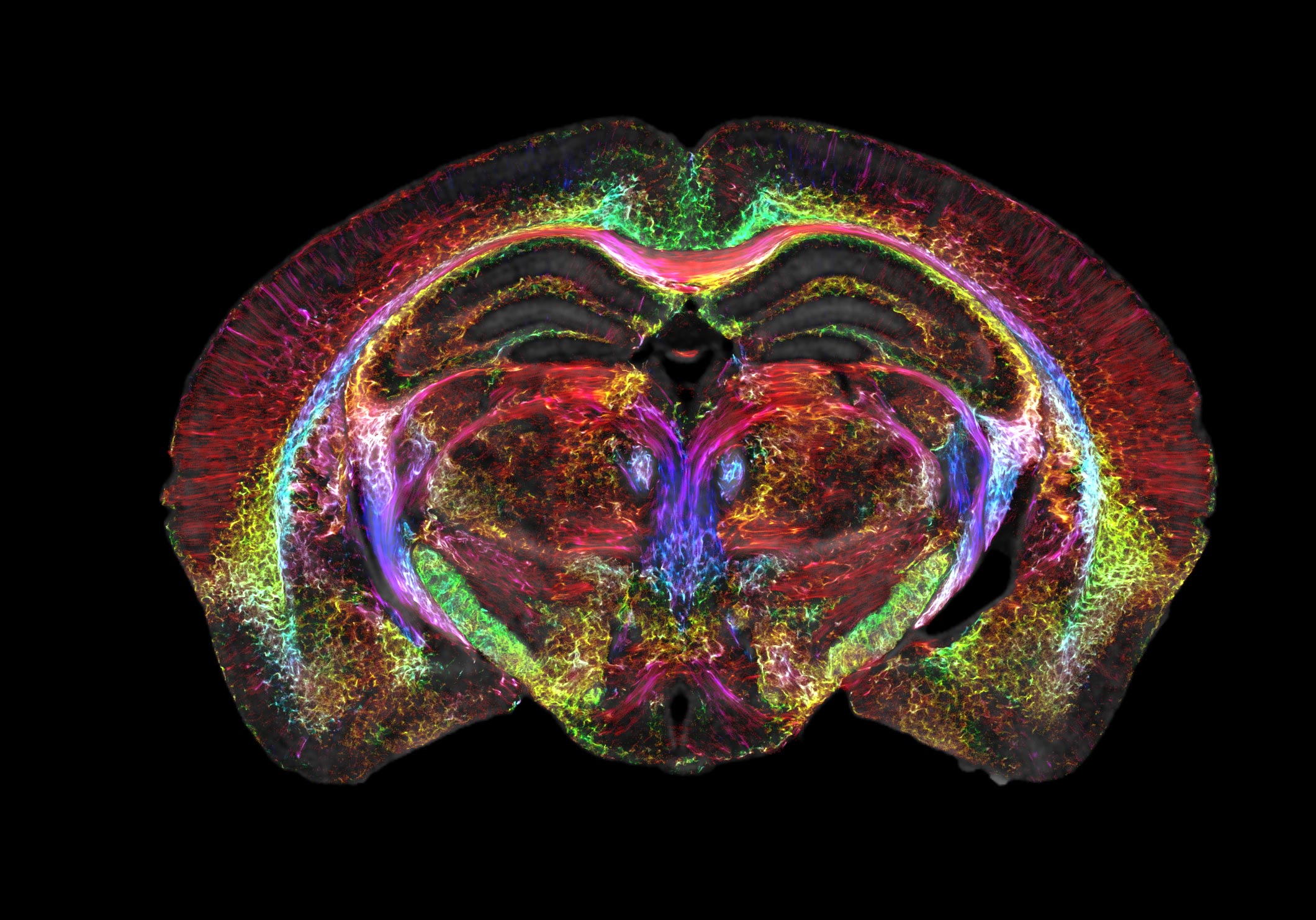
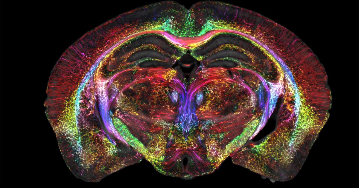
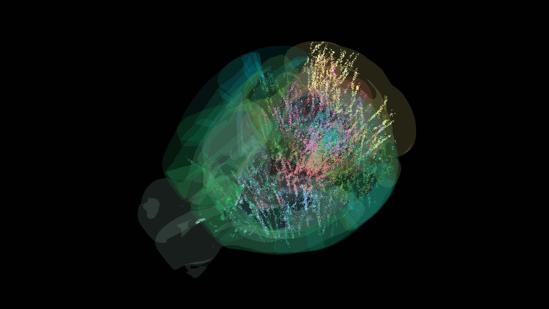
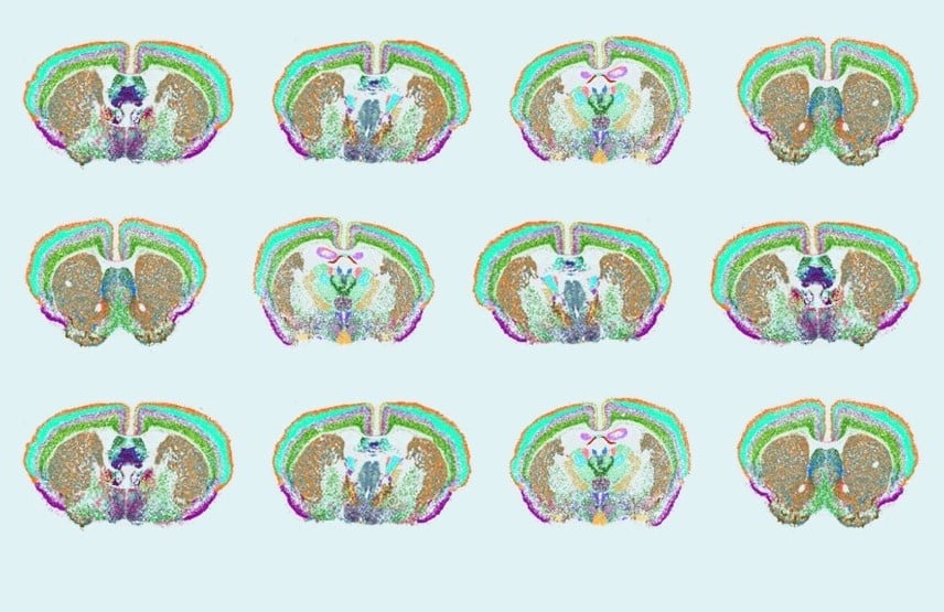
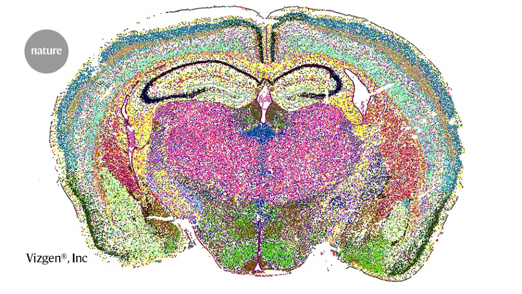
A groundbreaking international study mapped over 600,000 neurons across nearly the entire mouse brain, revealing that decision-making involves more brain regions than previously thought, challenging traditional models and highlighting the importance of collaborative, standardized research in neuroscience.
A new method utilizing a biocompatible nanosheet incorporated into light-curable resin (NIRE method) has been proposed to construct large cranial windows over the cortex and cerebellum suitable for long-term imaging in awake mice. The NIRE method produced windows that maintained transparency for over 5 months and suppressed motion artifacts, enabling the visualization of neural structures and intracellular Ca2+ concentration changes at various scales, from populations of over a thousand neurons to single spines, in living mouse brain. This method allows for multi-scale in vivo imaging of neuronal morphology and intracellular calcium in awake mice, demonstrating its potential for studying brain function and pathology.
Researchers have created the first complete cell atlas of a mammalian brain, specifically a mouse, providing detailed information on over 32 million cells, their types, locations, molecular profiles, and connectivity. This breakthrough offers a foundational map for understanding human brain functions and developing precision therapies for mental and neurological disorders. The atlas encompasses structural, transcriptomic, and epigenetic data, providing a blueprint for brain circuit operations and functioning. The findings were funded by the NIH BRAIN Initiative® and published in 10 papers in Nature.
Researchers have created a comprehensive atlas of the adult mouse brain, mapping the DNA methylome and 3D genome at single-cell resolution. The study utilized enhanced single-nucleus methylation sequencing and chromatin conformation capture sequencing to analyze DNA methylomes and the 3D genome in detail. The dataset identified 4,673 cell groups and provided a multi-omic resource for understanding the molecular architecture of the mouse brain. The study deepens insights into the epigenetic and transcriptomic intricacies that underpin brain function and diseases.
Scientists have created a complete cell atlas of a whole mammalian brain, specifically the mouse brain, providing detailed information on the type, location, and molecular characteristics of over 32 million cells. This atlas serves as a map for understanding the complex organization and connectivity of cells in the brain, paving the way for a better understanding of the human brain and the development of precision therapeutics for mental and neurological disorders. The findings were funded by the National Institutes of Health's BRAIN Initiative and published in Nature.
Researchers are making significant progress in mapping the mouse brain by combining high-throughput single-cell RNA sequencing with spatial transcriptomics. These methods allow for the identification and mapping of different categories of brain cells, providing comprehensive atlases of the mouse brain. The next steps involve understanding the functions of these molecularly defined cell types and creating a unified resource for the neuroscience community. These efforts are part of the larger BRAIN Initiative Cell Census Network, which aims to create comprehensive maps of cells in the brains of mice and primates, including humans.
Scientists have developed a high-resolution magnetic resonance imaging (MRI) technique that is 64 million times sharper than normal, allowing them to take high-definition images of a mouse brain. The technology could help detect changes to the human brain due to neurodegenerative diseases such as Alzheimer's disease and changes linked to healthy aging. The researchers used a high-powered 9.4-tesla magnet, gradient coils that are 100 times stronger than current models, and a high-speed computer to create the souped-up MRI. The technique could also be useful for studying how the brain changes when mice are put on specific diets or given drugs in an effort to extend their life spans.
Researchers from multiple universities have made a breakthrough in MRI technology, capturing the sharpest images ever of a mouse brain. This refined MRI, combined with light sheet microscopy, provides an unprecedented way to visualize the brain’s connectivity, potentially leading to a better understanding of neurodegenerative diseases in humans. The refined MRI provides an important new way to visualize the connectivity of the entire brain at record-breaking resolution. The researchers say new insights from mouse imaging will in turn lead to a better understanding of conditions in humans, such as how the brain changes with age, diet, or even with neurodegenerative diseases like Alzheimer’s.
Researchers have developed a new MRI imaging technology that dramatically improves the resolution of brain images, leading to the sharpest images ever generated of the mouse brain. The new images are 64 million times smaller than a clinical MRI voxel, allowing researchers to visualize microscopic details within the brain that reveal its organization. The refined MRI provides an important new way to visualize the connectivity of the entire brain at record-breaking resolution, leading to a better understanding of conditions in humans, such as how the brain changes with age, diet, or even with neurodegenerative diseases like Alzheimer’s.
Researchers from Duke University have developed a new MRI technology that provides the sharpest images ever captured of a mouse brain, with a single voxel measuring just 5 microns, 64 million times smaller than a clinical MRI voxel. The refined MRI provides an important new way to visualize the connectivity of the entire brain at record-breaking resolution, which will lead to a better understanding of conditions in humans, such as how the brain changes with age, diet, or even with neurodegenerative diseases like Alzheimer’s.