Single-cell proteomics maps human liver zonation and its fragility in diseased tissue
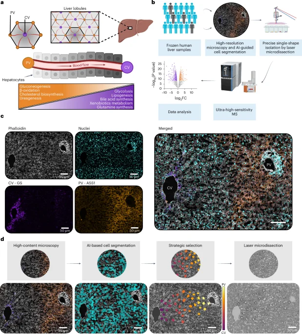
Researchers used advanced scDVP to profile hundreds of hepatocytes from 18 people, building a high-resolution map of protein gradients along the liver's porto–central axis. They quantified ~2,500 proteins per cell, showed roughly half are zonated, and introduced gradient-based analysis to quantify zonation without binning. Cross-species comparison with mice revealed shared and human-specific zonation features; in tissues with disrupted architecture, zonation is broadly lost. The study delivers an open-access liver proteome resource and a framework applicable to spatial proteomics.