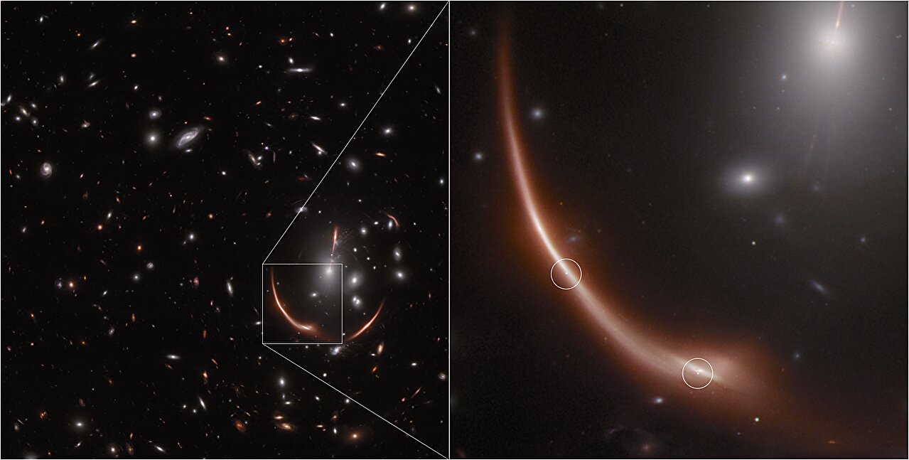
An international team has demonstrated that pixelized strong-lensing modeling on galaxy clusters significantly improves the precision of measuring the Hubble constant, potentially resolving the existing tension in its current measurements and advancing high-precision cosmology.
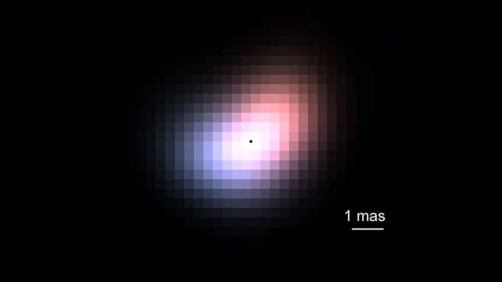
Astronomers have achieved the sharpest view of a distant star ever using a single telescope equipped with a novel device called a photonic lantern, which splits and analyzes starlight to produce high-resolution images, potentially revolutionizing the study of celestial objects.
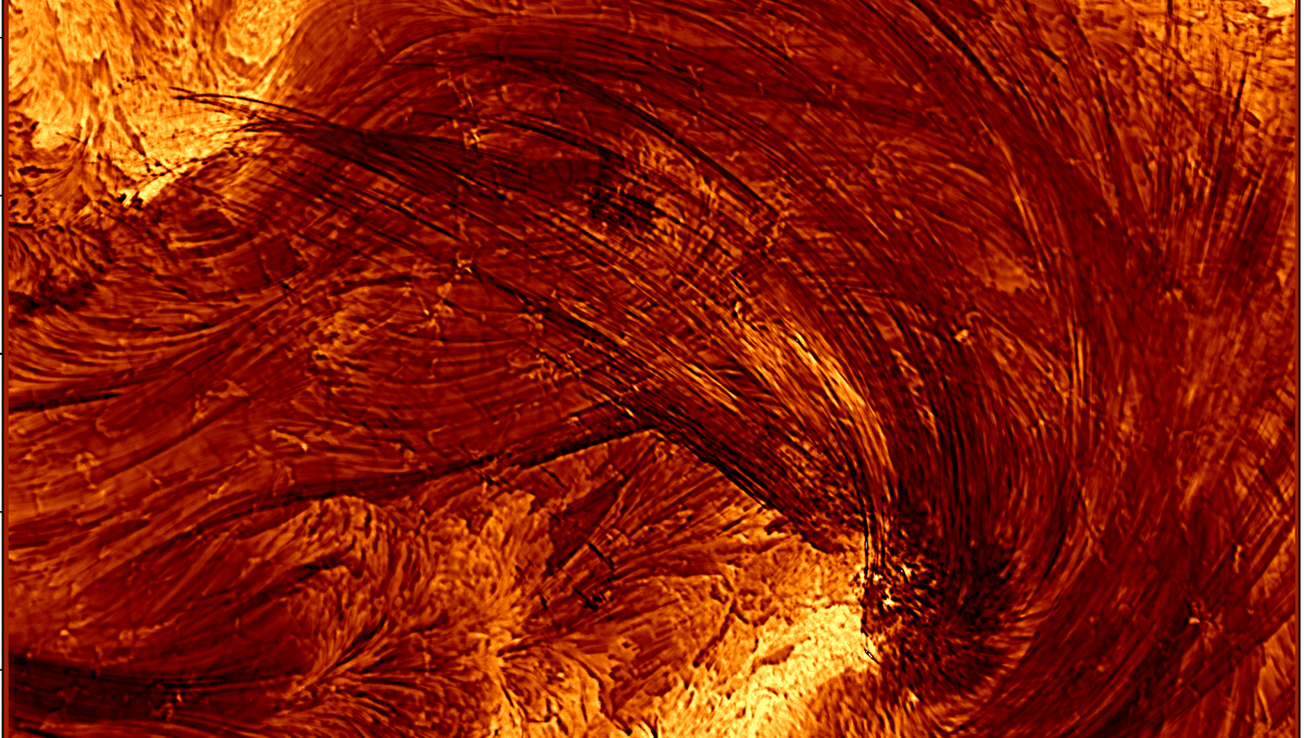
The Daniel K. Inouye Solar Telescope has captured the highest resolution images of coronal loops during a solar flare, revealing that these loops are much thinner than previously thought, with implications for understanding magnetic reconnection and solar activity.
Chinese scientists have developed a laser system capable of reading millimeter-sized text from nearly a mile away, surpassing traditional telescopic methods, with potential applications in archaeology and environmental monitoring, though it requires a clear line of sight and may incorporate AI for improved accuracy.
Sony has launched the A1 II camera, featuring a 50MP sensor, 30fps shooting, and advanced AI capabilities for automatic subject recognition. The camera boasts the fastest readout speed for non-global-shutter sensors, a 4-axis LCD monitor, and a noise reduction setting that combines multiple RAW files. It also includes a dedicated AI processing unit and a pre-capture feature. The A1 II will be available in December, alongside a new FE 28-70mm f/2 GM lens. The camera supports 2Gbps file transfer speed and has a body weight of 918g.
The James Webb Space Telescope has provided a stunning new high-definition look at the supernova remnant Cassiopeia A (Cas A) in the near-infrared spectrum. The image reveals intricate details of the expanding shell of material from the exploded star, including tiny knots of gas and light echoes. The image also highlights the absence of color in certain regions, indicating the presence of ionized gas and dust. Researchers are particularly intrigued by a large, striated blob called "Baby Cas A," which appears to be a light echo located about 170 light-years behind the supernova remnant.
Scientists at the National Institute of Standards and Technology (NIST) have developed a superconducting camera with 400,000 pixels, 400 times more than any previous device of its kind. The camera uses ultrathin electrical wires cooled to near absolute zero, where current moves with no resistance until a photon strikes a wire, disrupting the superconductivity and creating an electrical signal. By combining signals from multiple pixels onto a few readout wires, the researchers overcame the challenge of connecting each pixel to its own wire. This breakthrough could open up new applications in science, such as imaging faint galaxies or planets, measuring light in quantum computers, and biomedical studies using near-infrared light to examine human tissue.
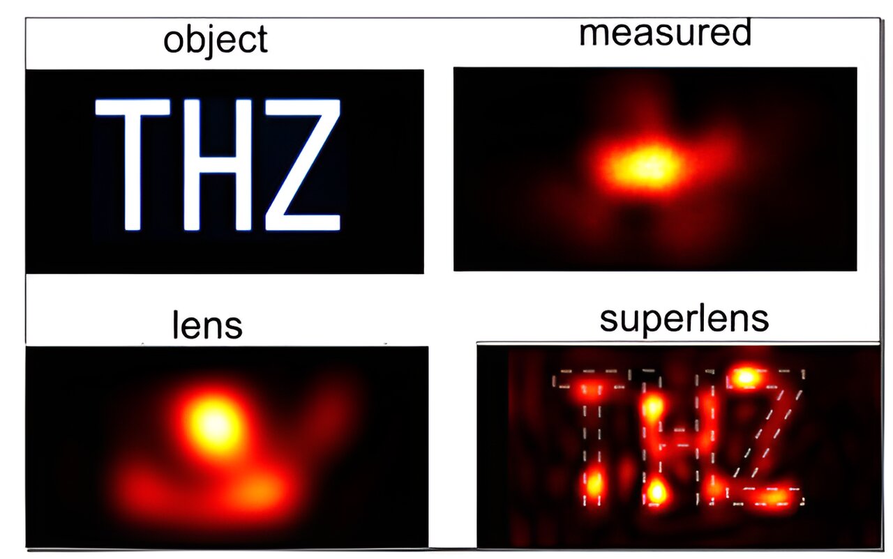
Researchers from the University of Sydney have developed a groundbreaking superlens technique that allows scientists to see objects four times smaller than the diffraction limit of traditional microscopes. By collecting high-resolution and low-resolution information using a light probe, and then selectively amplifying the valuable light waves through computer processing, the researchers were able to overcome the diffraction limit. This technique has potential applications in various fields, including medicine, diagnostics, microfabrication, and even art authentication. The researchers hope that their method will enable high-resolution imaging while maintaining a safe distance from the object being observed.
Physicists at the University of Sydney have developed a new technique called superlensing that allows for high-resolution imaging beyond the diffraction limit without the need for a super lens. By placing the light probe far away from the object and collecting both high- and low-resolution information, they were able to selectively amplify evanescent light waves to produce a clear image. This breakthrough could have applications in fields such as cancer diagnostics, medical imaging, archaeology, and forensics.
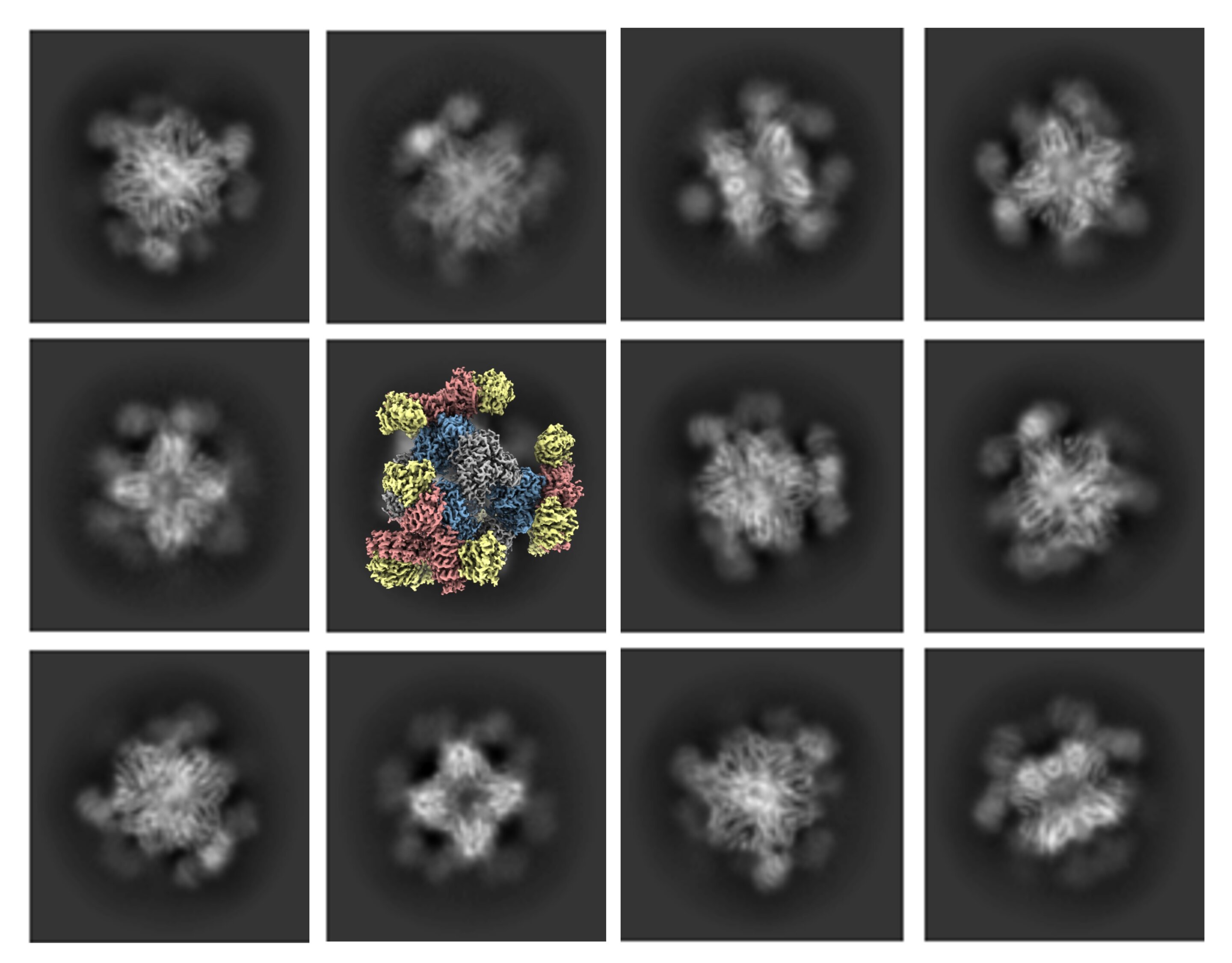
Scientists at UCLA have developed a solution to improve cryo-electron microscopy (cryo-EM) by enabling high-quality imaging of smaller protein molecules. They engineered a cube-shaped protein structure called a scaffold with tripod-like protrusions that hold the small proteins in place. The scaffold can be digitally removed during image processing, resulting in a composite 3D image of the small protein being analyzed. This advancement expands cryo-EM's imaging capabilities and has potential applications in drug development, allowing researchers to identify specific locations on proteins for therapeutic targeting. The technique was successfully tested on a protein involved in cancer treatments.
Researchers have developed a complex-domain neural network that enhances large-scale coherent imaging, revolutionizing optical imaging by providing wide field-of-view and high-resolution capabilities. The technique exploits latent coupling information between amplitude and phase components, leading to multidimensional representations of complex wavefront. The network significantly reduces exposure time and data volume while maintaining high-quality reconstructions, offering implications for high-level semantic analysis and intelligent medical care. This technology holds promise for real-time cell observation and pushing the boundaries of medical diagnostics.
The European Space Agency has released a stunning photo of Earth captured by its latest satellite imager, the Meteosat Third Generation Imager-1 (MTG-I1), which can produce imagery at a much higher resolution and more frequently than its predecessors. The image reveals weather conditions above Africa, Europe, and the Atlantic, with details such as cloud vortices over the Canary Islands, snow cover on the Alps, and sediment in the water along the coast of Italy now visible to scientists. The MTG-I1 is currently undergoing a 12-month calibration phase, and its high-resolution imaging is expected to revolutionize weather forecasting.
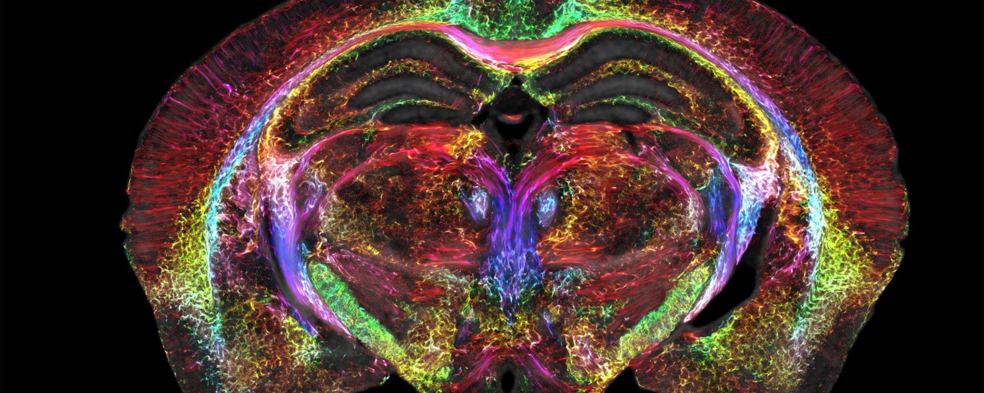
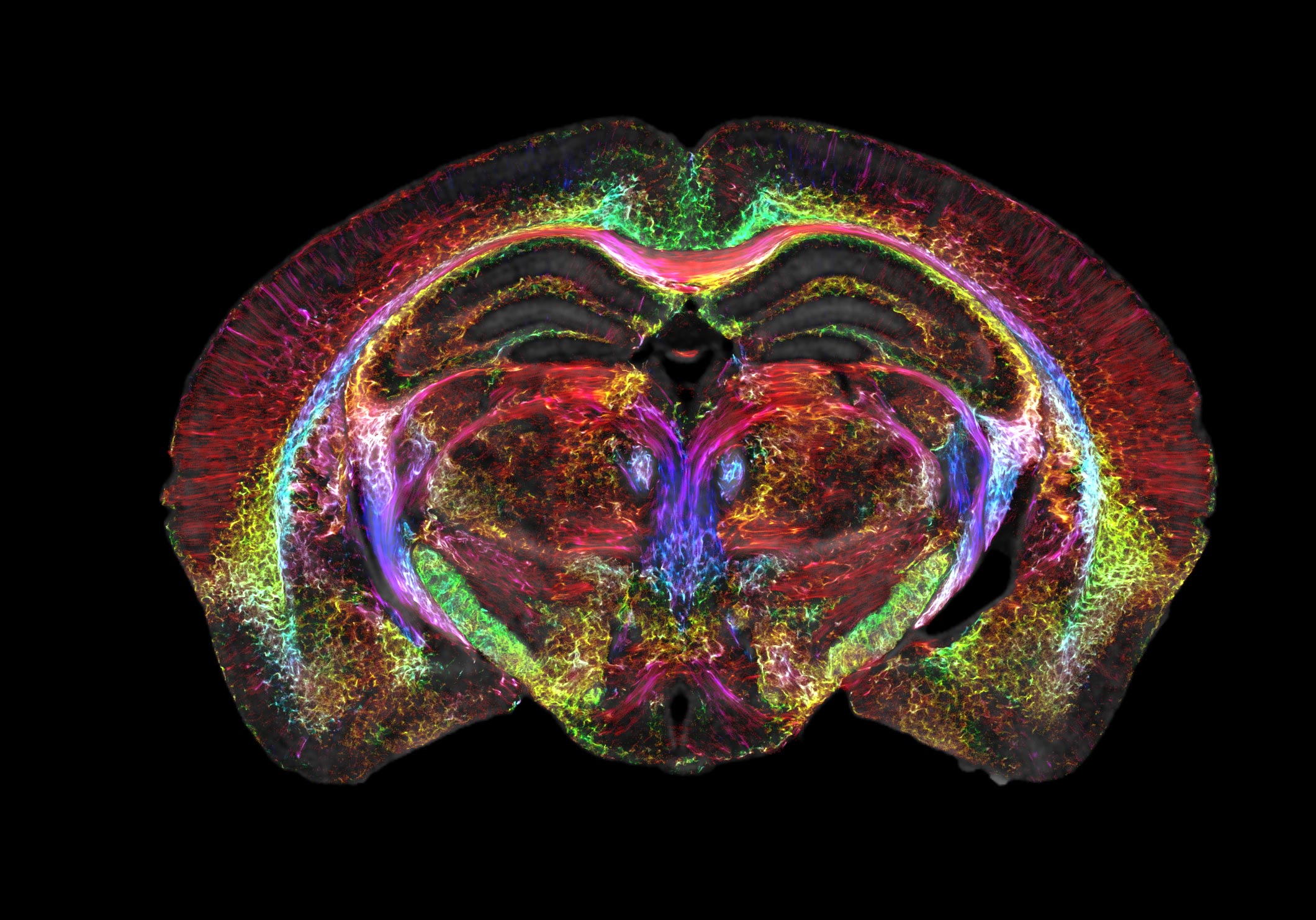
Duke University researchers have developed a new MRI technology using a 9.4 Tesla magnet and specialized gradient coils, leading to an image resolution six orders of magnitude higher than a typical MRI. The voxels in the image measure at only 5 microns, revealing microscopic details within brain tissues that were previously unattainable. This breakthrough in MRI resolution has the potential to significantly advance understanding of the neural networks found in humans by first studying neural structures in mice at this unprecedented detail.
Researchers from multiple universities have made a breakthrough in MRI technology, capturing the sharpest images ever of a mouse brain. This refined MRI, combined with light sheet microscopy, provides an unprecedented way to visualize the brain’s connectivity, potentially leading to a better understanding of neurodegenerative diseases in humans. The refined MRI provides an important new way to visualize the connectivity of the entire brain at record-breaking resolution. The researchers say new insights from mouse imaging will in turn lead to a better understanding of conditions in humans, such as how the brain changes with age, diet, or even with neurodegenerative diseases like Alzheimer’s.
Researchers have developed a new MRI imaging technology that dramatically improves the resolution of brain images, leading to the sharpest images ever generated of the mouse brain. The new images are 64 million times smaller than a clinical MRI voxel, allowing researchers to visualize microscopic details within the brain that reveal its organization. The refined MRI provides an important new way to visualize the connectivity of the entire brain at record-breaking resolution, leading to a better understanding of conditions in humans, such as how the brain changes with age, diet, or even with neurodegenerative diseases like Alzheimer’s.