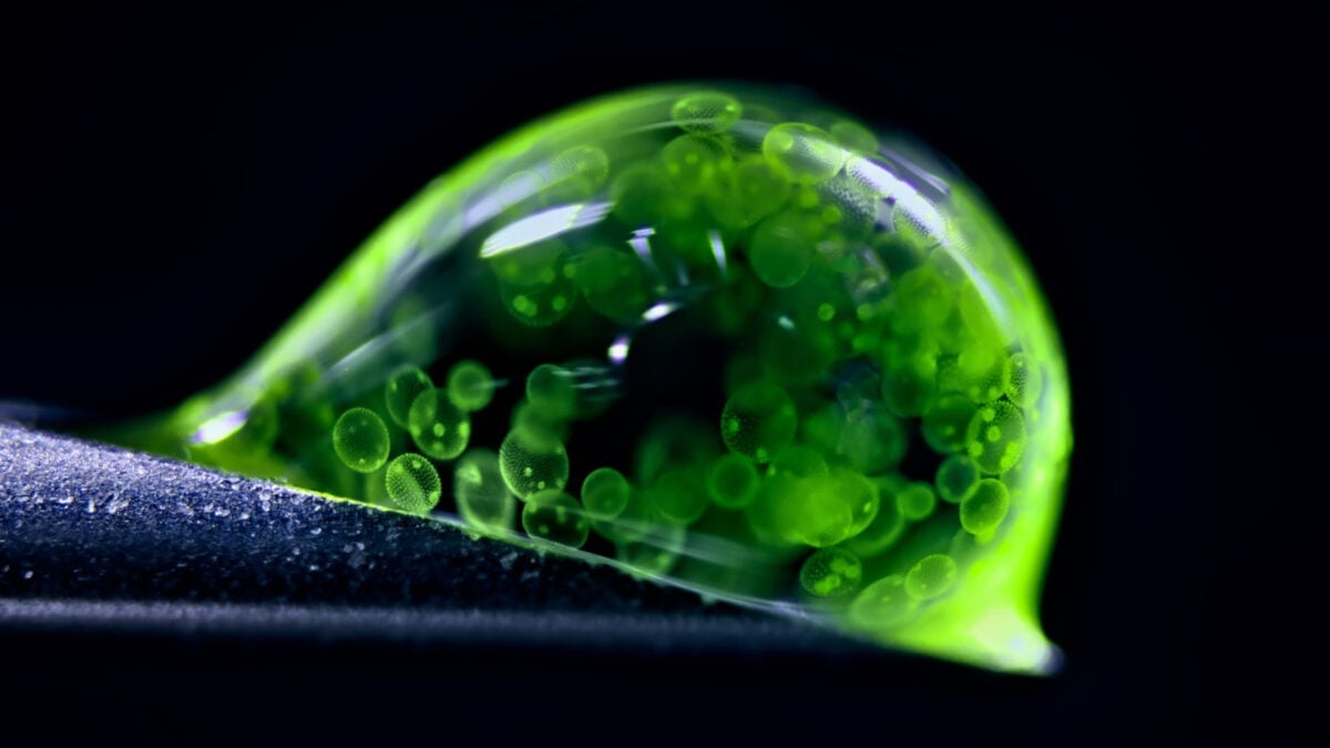
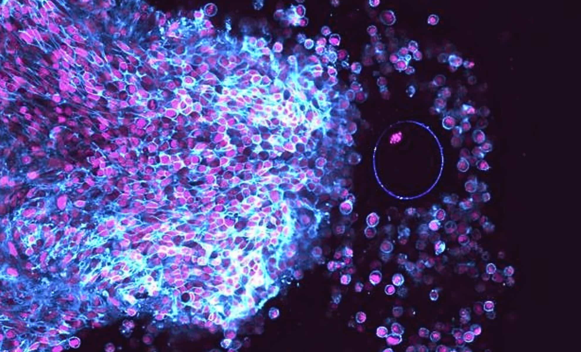
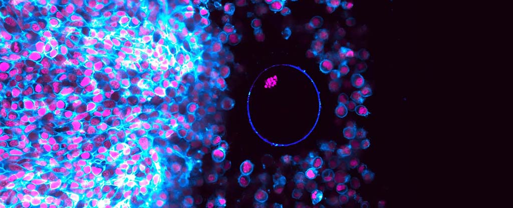
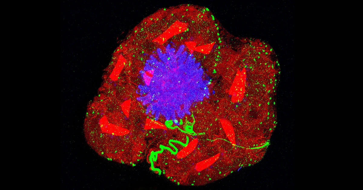
Tag

Microscopy

All articles tagged with #microscopy

Nikon Small World 2025: Stunning Microphotography Winners Unveiled

Originally Published 2 months ago — by The Washington Post

Featured image for Nikon Small World 2025: Stunning Microphotography Winners Unveiled
Source: The Washington Post

The 51st Nikon Small World Photomicrography Competition recognized 71 outstanding images captured through microscopy, with China's Zhang You winning first place for his detailed photo of a rice weevil on rice, highlighting the intricate world of insects and their ecological roles.

Nikon Small World 2025: Stunning Microscopic Images Unveiled

Originally Published 2 months ago — by ScienceAlert

Featured image for Nikon Small World 2025: Stunning Microscopic Images Unveiled
Source: ScienceAlert

The article highlights award-winning microscopic images from the 2025 Nikon Small World competition, showcasing intricate details of tiny life forms like insects, cells, and microorganisms, emphasizing the importance of understanding and appreciating the complexity of microscopic worlds.

Scientists Develop the World's Smallest Violin Using Nanotechnology

Originally Published 7 months ago — by Newser

Featured image for Scientists Develop the World's Smallest Violin Using Nanotechnology
Source: Newser

Scientists at Loughborough University have created the world's smallest violin, measuring just 35 by 13 microns, as a demonstration of their nanolithography capabilities, which could pave the way for advancements in computing and energy harvesting, though the violin is not playable.

The Power of Curiosity and Paradox in Scientific Discovery

Originally Published 7 months ago — by Quanta Magazine

Featured image for The Power of Curiosity and Paradox in Scientific Discovery
Source: Quanta Magazine

Manu Prakash, a Stanford bioengineer, advocates for a philosophy called 'recreational biology,' which emphasizes curiosity and wonder in scientific exploration. He is renowned for creating low-cost scientific tools like the Foldscope, a paper microscope, and for studying both urgent health issues and the fascinating behaviors of microscopic organisms, blending practical applications with fundamental science to inspire broad curiosity and democratize access to scientific discovery.

Real-Time Ovulation Captured: A Breakthrough for Infertility Treatment?

Originally Published 1 year ago — by The Daily Galaxy --Great Discoveries Channel

Featured image for Real-Time Ovulation Captured: A Breakthrough for Infertility Treatment?
Source: The Daily Galaxy --Great Discoveries Channel

Researchers at the Max Planck Institute have captured the first real-time video of ovulation, revealing the process in unprecedented detail. This breakthrough, achieved through advanced microscopy and live tissue culture techniques, allows scientists to observe the three key phases of ovulation, potentially revolutionizing infertility treatments. By understanding ovulation more precisely, this discovery could lead to improved diagnostics and treatments for conditions like polycystic ovary syndrome (PCOS), marking a significant advancement in reproductive medicine.

First-Ever Footage Captures Complete Ovulation Process

Originally Published 1 year ago — by ScienceAlert

Featured image for First-Ever Footage Captures Complete Ovulation Process
Source: ScienceAlert

Scientists at the Max Planck Institute have successfully filmed the entire ovulation process in mice for the first time, using advanced microscopy techniques. This breakthrough allows researchers to observe the intricate dynamics of ovulation in unprecedented detail, potentially aiding in understanding conditions like polycystic ovary syndrome (PCOS) where ovulation is impaired. The study, published in Nature Cell Biology, marks a significant advancement in reproductive research.

Breakthrough Microscope Captures Nanoscale Biology in Real-Time

Originally Published 1 year ago — by Phys.org

Featured image for Breakthrough Microscope Captures Nanoscale Biology in Real-Time
Source: Phys.org

Researchers at Radboud University Medical Center in the Netherlands have developed a groundbreaking microscope that allows for live imaging of biological processes at the nanoscale, overcoming previous limitations of either frozen or low-detail observations. This technique uses graphene to protect samples from electron beam damage, enabling detailed visualization of processes like arterial calcification. The innovation could lead to new insights into diseases and treatments, such as developing a 'heart valve on a chip' to study calcification.

"Scientists Unveil X-Ray Vision to Peer Inside Crystals"

Originally Published 1 year ago — by Phys.org

Featured image for "Scientists Unveil X-Ray Vision to Peer Inside Crystals"
Source: Phys.org

Researchers at New York University have developed a technique called "Crystal Clear" that allows scientists to visualize the internal structure of colloidal crystals in three dimensions. Using transparent particles, dye molecules, and confocal microscopy, they can create dynamic models to study the arrangement and defects within crystals, providing new insights into crystallization processes and potentially aiding in the development of better materials.

"Scientists Unveil Secret Behind Butterfly Wing Colors"

Originally Published 1 year ago — by Good News Network

Featured image for "Scientists Unveil Secret Behind Butterfly Wing Colors"
Source: Good News Network

Researchers at the University of Sheffield and the Central Laser Facility have discovered that the protein actin is crucial in forming the vibrant colors of butterfly wings by orchestrating the arrangement of colorful structures in the scales. Using high-resolution microscopy, they observed that denser actin bundles create more reflective ridges, and altering these structures can cause colors to fade. This discovery could inspire new technologies in sensing, diagnostics, and sustainable paints. The study was published in Nature Communications.

"Breakthrough: Resolving Decades-Long Microscopy Problem"

Originally Published 1 year ago — by Phys.org

Featured image for "Breakthrough: Resolving Decades-Long Microscopy Problem"
Source: Phys.org

A research team has resolved a long-standing issue in microscopy by developing a depth-dependent scaling factor to correct the flattening effect caused by different mediums in the lens and sample. This breakthrough allows for more precise imaging of biological structures, potentially aiding in the study and understanding of abnormalities and diseases. The team has provided a web tool and software for determining the corrective factor, which can significantly reduce time and costs associated with microscopy experiments.

"Revolutionizing Cell Exploration: Advanced Microscopy Unveils Intricate Details"

Originally Published 1 year ago — by Phys.org

Featured image for "Revolutionizing Cell Exploration: Advanced Microscopy Unveils Intricate Details"
Source: Phys.org

Yale scientists have developed a new microscopy technique called FLASH-PAINT, which allows researchers to view an unlimited number of different molecules within individual cells. This technique, published in Cell, utilizes imaging probes and an adapter that transiently binds to numerous targets, making it 100 times faster and more cost-efficient than current super-resolution microscopy techniques. The team hopes that FLASH-PAINT will enable visualization of complex subcellular processes, leading to new insights for disease treatment and potential diagnostic applications.

"First-ever Reconstruction of Human Centriole Assembly Image by Image"

Originally Published 1 year ago — by Phys.org

Featured image for "First-ever Reconstruction of Human Centriole Assembly Image by Image"
Source: Phys.org

Scientists at the University of Geneva have used high resolution microscopy and kinematic reconstruction techniques to visualize the assembly of the human centriole, a crucial organelle in cell organization. By combining expansion microscopy and kinematic reconstruction, they reconstructed the first 4D assembly of the human centriole, shedding light on its formation and opening new possibilities for studying other cellular structures.