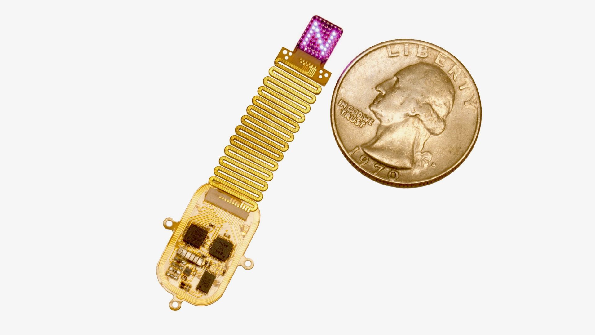
A small, wireless implant uses LED light and genetic modification to communicate directly with the brain's neurons in mice, enabling new ways to study and potentially treat neurological conditions without invasive procedures or external wires.
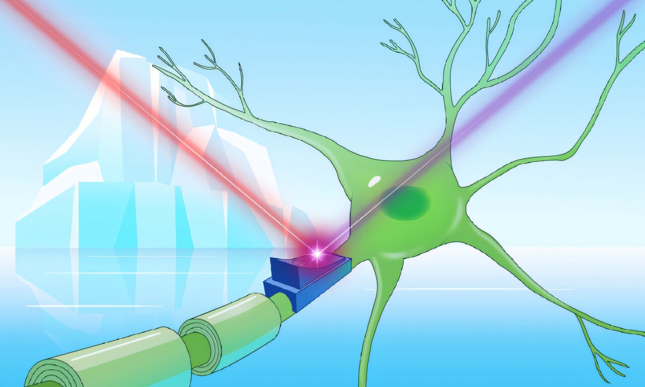
Researchers discovered unique blue cryorhodopsins in cold-environment microbes that can sense UV light and control cellular activity, offering potential for advanced optogenetic tools and insights into microbial adaptation to UV exposure in icy habitats.
A rat study suggests that stimulating the brain region involved in sound and emotion processing, the inferior colliculus, may help alleviate motor symptoms of Parkinson's disease without affecting emotional responses, potentially opening new avenues for treatment beyond traditional basal ganglia targets.
Researchers have discovered that progesterone-responsive neurons in the anterior ventromedial hypothalamus (VMH) of female mice toggle between sexual receptivity and rejection based on fertility. These neurons are active during rejection behaviors outside the fertile phase and receive inhibitory signals during fertility, reducing their activity and allowing mating. Using optogenetics, the study confirmed these neurons act as a neural switch for rejection, offering insights into human sexual behavior and related disorders.
Researchers at UC Davis have identified distinct neural circuits responsible for the anti-anxiety effects of psychedelics, separate from those causing hallucinations. Using the psychedelic DOI in mice, they found that anxiety reduction persists after hallucinatory effects fade. By mapping and reactivating specific neurons in the prefrontal cortex, they demonstrated potential for developing psychedelics-based treatments that alleviate anxiety without inducing hallucinations. This study highlights the complexity of psychedelic effects, involving both direct and downstream neural networks.
Researchers at Tohoku University have discovered that astrocytes, cells surrounding neurons, play a crucial role in determining which memories are retained or forgotten. By using optogenetics to manipulate astrocytes in mice, they found that acidifying these cells after a traumatic event leads to memory loss, while alkalinizing them preserves memories long-term. This finding challenges the traditional view that short- and long-term memories form sequentially, suggesting they may develop in parallel. The study could inform new treatments for PTSD by targeting astrocyte functions.
Researchers at OIST and Keio University have discovered that stimulating the brain's dorsal raphe nucleus (DRN), the main source of serotonin, activates areas responsible for behavior and motivation in awake mice. Using opto-functional MRI, they observed that DRN serotonin activation stimulates the cerebral cortex and basal ganglia, providing insights into serotonin's role in mood regulation and cognitive functions. This study could advance understanding of mood disorders and behavioral adaptations.
A study using optogenetics and high-field MRI on awake mice reveals that activating serotonin neurons in the dorsal raphe nucleus stimulates the cerebral cortex and basal ganglia, impacting behavior and motivation. This research enhances understanding of serotonin's role in brain-wide activation and its implications for mood therapy and behavioral adaptation.
MIT researchers have developed an optogenetic technique to control muscles using light, offering more precise control and significantly reducing fatigue in mice compared to traditional electrical stimulation. This approach, while not yet feasible in humans, could revolutionize prosthetics and aid individuals with impaired limb function. The team is working on safely delivering light-sensitive proteins to human tissue to make this method clinically viable.
MIT researchers have developed an optogenetic technique to control muscle contractions using light, offering more precise control and reduced fatigue compared to traditional electrical stimulation. This method, tested in mice, could potentially benefit people with paralysis, amputations, and other limb impairments, though challenges remain in safely delivering light-sensitive proteins to human tissue.
A study published in Neuropsychopharmacology Reports reveals the potential of delta opioid receptors to alleviate anxiety through the activation of a specific brain pathway. The selective DOP agonist, KNT-127, was found to reduce anxiety-like behavior in mice, offering promise for developing new treatments for anxiety-related disorders. The study's methodology, anchored in optogenetics, targeted a neural pathway from the prelimbic cortex to the basolateral nucleus of the amygdala, critical areas associated with emotion regulation and anxiety development. The findings underscore the pivotal role of this brain circuit in regulating innate anxiety and demonstrate the potential of DOP agonists, like KNT-127, in alleviating anxiety.
A new study using optogenetics has revealed that the perception of time is intertwined with the sense of touch, with the somatosensory cortex playing a dual role in processing both tactile sensations and time perception. The research demonstrates that the perception of time is rooted in a widespread network of brain areas, shedding light on the intricate interplay between the sense of touch and the sense of time. This insight opens new avenues for understanding the complex relationship between sensing the external world and sensing time.
A groundbreaking study published in Nature Communications reveals a link between the sense of touch and time perception, demonstrating the crucial role of the somatosensory cortex in how we perceive the duration of tactile experiences. Utilizing optogenetics, researchers found that increasing neuronal activity in this brain region led to altered perceptions of both intensity and duration of tactile stimuli in rats. The study challenges the traditional view of time perception and suggests an integrated approach to understanding sensory experiences, while also providing a theoretical framework for linking neural processes to subjective experiences.
A new study suggests that the superior colliculus, a small pea-sized region in the human brain, plays a more significant role than previously thought, alongside the visual cortex, in how mice perceive their immediate surroundings. Researchers from the Netherlands Institute for Neuroscience used optogenetics to switch off the superior colliculus in mice and found that it significantly impaired their ability to detect objects. The study indicates that this tiny brain region may be more important than previously believed and could be responsible for visual perception in both mice and humans.
Schwann cells, traditionally known for insulating nerve fibers, have been discovered to play a crucial role in detecting sensory stimuli such as touch and pain. This groundbreaking study utilized optogenetics to manipulate these cells in mice, demonstrating their significant role in transmitting pain sensations and potential as a novel target for pain therapy. The findings challenge the existing understanding of sensory perception and offer promising new directions for treating pain and tactile impairments.