Headache Misread as Stress Uncovers Tennis-Ball Brain Tumor
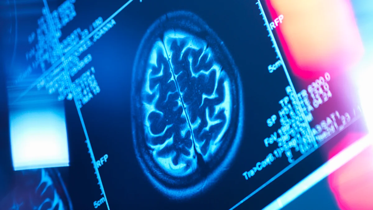
A 21-year-old college student’s severe headaches were initially dismissed as stress during finals, but an MRI revealed a two-inch benign brain tumor pressing on the left frontal cortex. She underwent a six-hour craniotomy to remove it and is now preparing for radiation therapy to prevent regrowth, urging others to trust their gut and seek second opinions if symptoms persist.