Scientists have created embryo-like structures from stem cells in the lab that can produce human blood cells, opening new avenues for regenerative medicine and understanding early human development, with potential applications in treating blood disorders and customizing therapies using a patient's own cells.
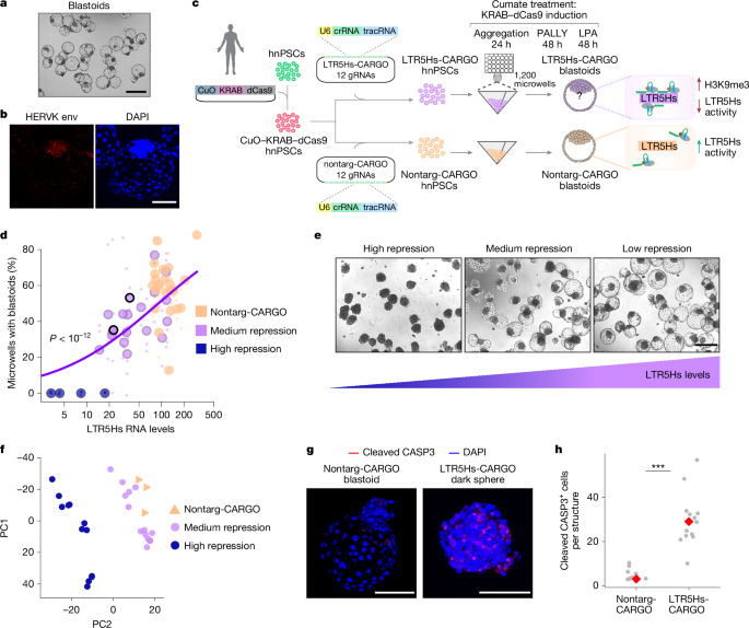
The study uncovers a human-specific regulatory mechanism involving endogenous retroviruses (ERVs), particularly HERVK LTR5Hs, which influence gene expression and lineage specification during early human development, using a stem cell-based blastoid model. Repression of LTR5Hs impairs blastoid formation, alters lineage allocation, and affects the expression of key genes like ZNF729, a human-specific gene regulated by a nearby LTR5Hs insertion that is essential for blastoid formation and proliferation. The work highlights the evolutionary role of ERVs as enhancers shaping human-specific developmental features.
Scientists have for the first time filmed the dynamic process of human embryo implantation in a lab setting, providing new insights into early development and potential improvements in fertility treatments.
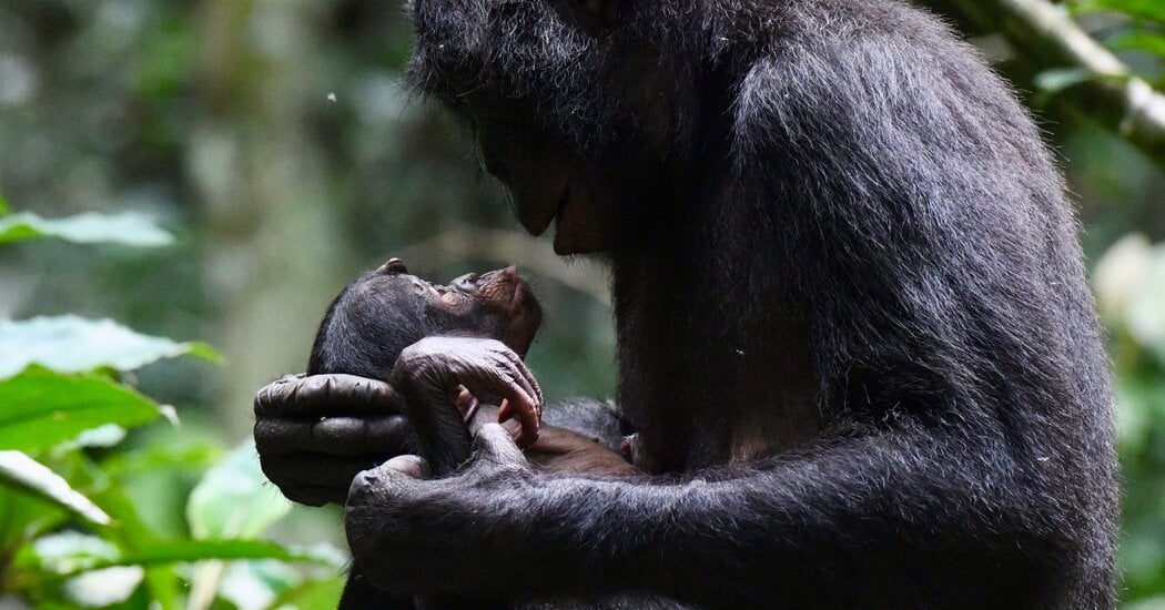
A study comparing communication between human children and apes suggests that the unique and frequent infant-directed speech in humans may have played a crucial role in the evolution of language, with humans engaging in much more frequent and elaborate communication with their young than apes do.
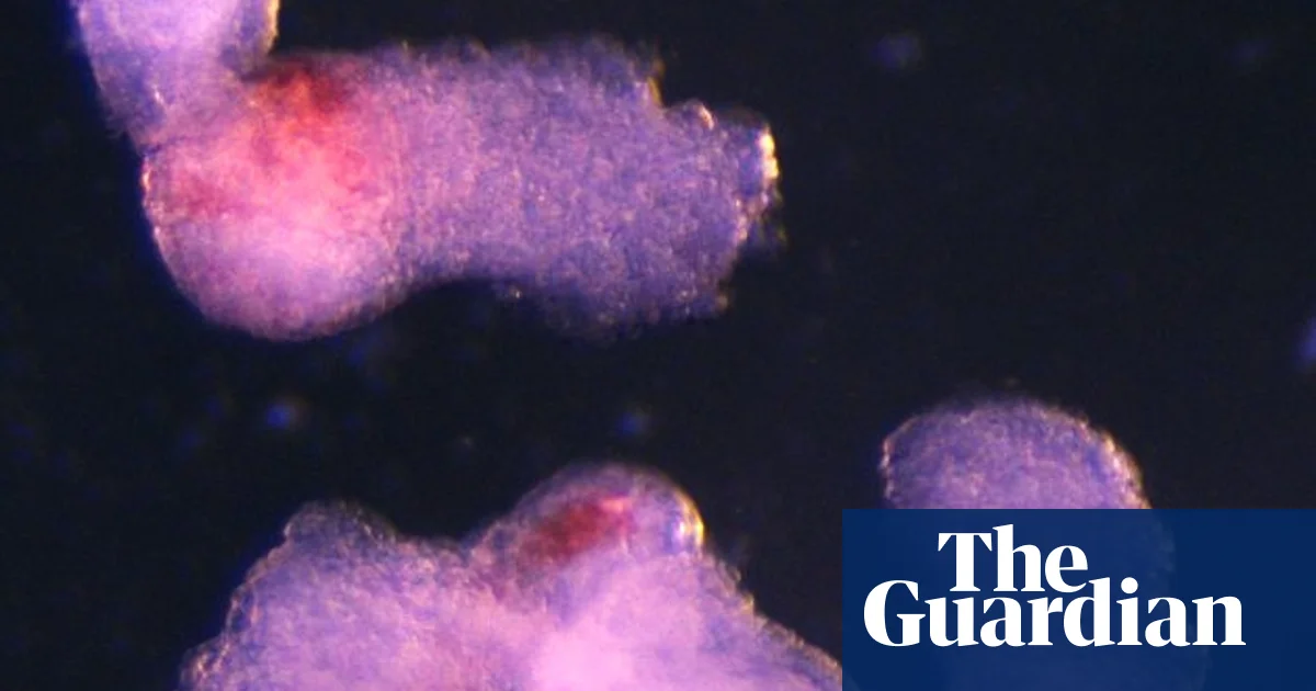
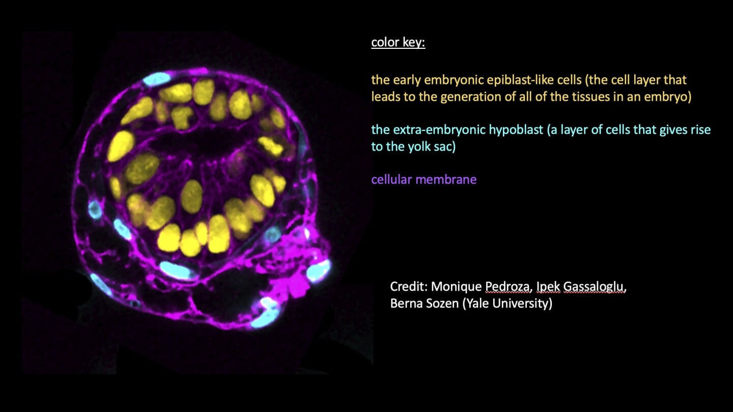
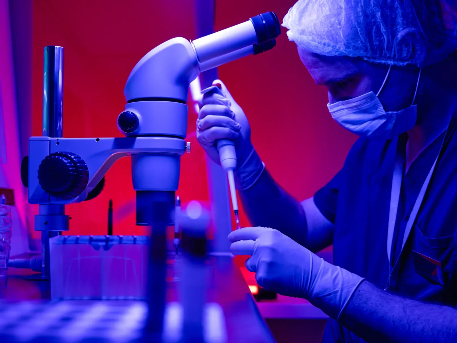
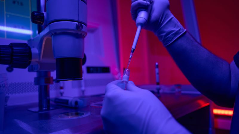
Researchers have developed a genetically inducible stem cell-derived embryoid model that mimics early post-implantation human embryogenesis. This model captures the co-development of embryonic tissue and extra-embryonic endoderm and mesoderm niche, as well as the emergence of hematopoiesis. The embryoids exhibit self-organizing cellular programs similar to those seen in embryogenesis, including the formation of amniotic cavity and bilaminar disc morphologies. The extra-embryonic layer in these embryoids undergoes yolk sac tissue-like morphogenesis and shows distinct waves of hematopoiesis. This model provides a scalable platform for studying human development and blood formation, and can be used for drug testing and disease modeling.
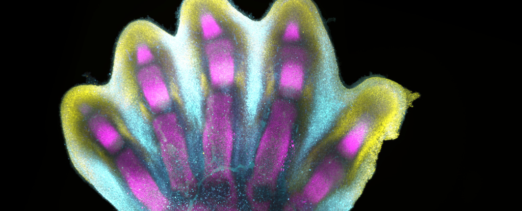
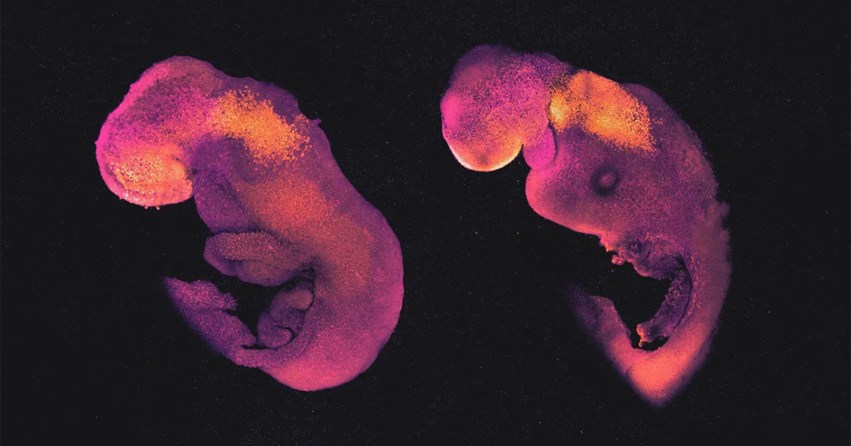
Scientists have created the first human cell atlas of early limb development, revealing in exquisite detail how fingers and toes grow. By analyzing thousands of single cells from donated embryonic tissues, the researchers mapped gene expression patterns and identified distinct cell clusters involved in limb development. They found that the process is highly complex and precisely regulated, resembling a sculptor chiseling away at a block of marble. The study deepens our understanding of how anatomically complex structures form and has implications for research and healthcare. The researchers also showed that limb formation in humans and mice follows similar trajectories, with some differences in activated genes and cell types.
A study conducted by Florida Atlantic University (FAU) has shed light on the origins of agency and conscious awareness by observing the behavior of human infants. By tethering an infant's foot to a mobile, researchers found that each foot movement caused the mobile to move, creating a positive feedback loop that highlighted the cause-and-effect relationship between the infant and the mobile. This realization led to a transition from spontaneous to intentional behavior, marked by an abrupt increase in infant movement rate. The study also revealed that agency emerges from the coordinated relationship between the infant and the environment. The findings provide valuable insights into the development of conscious awareness in humans.
Israeli scientists at the Weizmann Institute have achieved a significant breakthrough in synthetic embryo development by creating a stem cell-derived human embryo model that closely resembles a human embryo at day 14 of development. This advancement will enable researchers to study the development of organs, as well as investigate birth defects and congenital diseases that occur during the critical period between day 10 and day 40 of embryonic development.
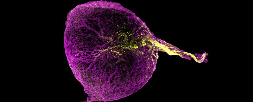
Researchers from the Wellcome Sanger Institute have discovered that the seemingly useless yolk sac in human embryos actually plays a crucial role in the development of the immune system. By sequencing RNA strands from human yolk sac cells, the researchers created a detailed atlas of the tissues, revealing that the yolk sac is responsible for producing the first blood cells and contributing to various important early functions. The study also identified significant differences between human yolk sac tissues and those in typical lab models, providing valuable insights into early human development and potential applications in disease research and tissue engineering.
Scientists have created embryo models using stem cells to study early human development and understand medical problems that occur before birth. These models, which resemble embryos after implantation in the uterus, provide insights into the hidden stages of human development. Researchers from the United States, England, Israel, and China have published studies on their work, describing models that mimic human embryos up to 14 days after fertilization. These models can help study embryonic failure, developmental disorders, and pregnancy loss, as well as explore the effects of the environment and chemicals on early development. However, ethical guidelines prevent the use of these models for reproduction, and they are not subject to the "14-day rule" that limits the growth of actual embryos in the lab.
Researchers have developed a system using human pluripotent stem cells that can self-organize into three-dimensional structures that mimic key events of early human post-implantation embryonic development. The system captures spontaneous differentiation and co-development of embryonic epiblast and extra-embryonic hypoblast-like lineages, establishes key signaling hubs with secreted modulators, and can undergo symmetry breaking-like events. Single-cell transcriptomics confirms differentiation into diverse cell states of the peri-gastrulating human embryo without establishing placental cell types, offering a reproducible, tractable, and scalable experimental platform to understand the basic cellular and molecular mechanisms that underlie human development.
Scientists have created the first synthetic human embryos using stem cells, which could help researchers study the earliest stages of human development and explain pregnancy loss. However, the rapid progress has outpaced discussions on how they should be dealt with ethically and legally. The synthetic embryos are not legally "embryos" and are not governed by the same laws as traditional embryos. Legal and ethical experts in the UK are drawing up a voluntary set of guidelines for how to proceed.
Scientists have created the world's first human synthetic embryos from stem cells without using sperm or eggs. These embryo-like structures lack organs such as a beating heart or a brain, but include cells that would typically go on to form the placenta, yolk sac and the embryo itself. The research raises legal and ethical questions, as many countries currently lack regulations looking at the creation and manipulation of synthetic embryos. There is an urgent need for regulations to provide a framework for the creation and use of stem cell-derived models of human embryos.
Scientists in the US and UK have created synthetic human embryo-like structures from stem cells, which could help advance the understanding of genetic diseases or the causes of miscarriages. The embryo-like structures were grown from single human embryonic stem cells that were coaxed to develop into three distinct tissue layers, including cells that would typically go on to develop a yolk sac, a placenta and the embryo itself. The research raises critical legal and ethical questions, and many countries, including the US, don’t have laws governing the creation or treatment of synthetic embryos.
Embryo models made from embryonic stem cells can self-organize into a hollow form shaped like a peanut shell and comparable in appearance to a regular embryo undergoing gastrulation. These models can be used to experimentally study human development and perhaps lead to a better understanding of defects that cause miscarriages or deformities. However, the ethical and biological boundaries become blurry as the models get better and further along. The failure rate for growing embryo models is very high, and more work is needed to understand both the similarities to normal embryos and the differences that may explain why mouse embryo models haven’t been able to grow beyond 8.5 days.