Researchers have developed a novel X-ray technique called stochastic Stimulated X-ray Raman Scattering (s-SXRS) that uses noise to achieve unprecedented resolution in atomic and electronic structure imaging, enabling detailed insights into chemical reactions and material properties, with potential widespread applications in science and industry.
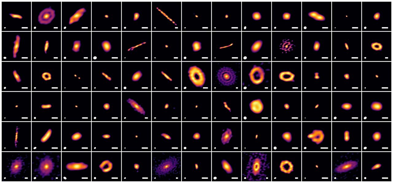
New super-resolution imaging of protoplanetary disks in the Ophiuchus region reveals that characteristic structures indicating planet formation appear within a few hundred thousand years after star birth, suggesting planets begin forming much earlier than previously thought. The study used advanced imaging techniques to analyze 78 disks, finding that planets grow alongside their young stars, with structures emerging in disks larger than 30 au during early star formation stages.
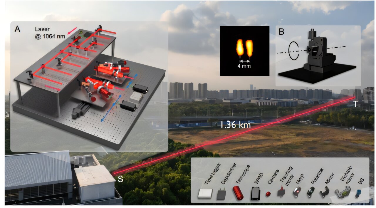
Researchers have developed a high-resolution laser device using intensity interferometry that can read millimeter-scale text from over a mile away, demonstrating super-resolution imaging at 1.36 km in an outdoor environment, with potential applications in remote sensing and surveillance.
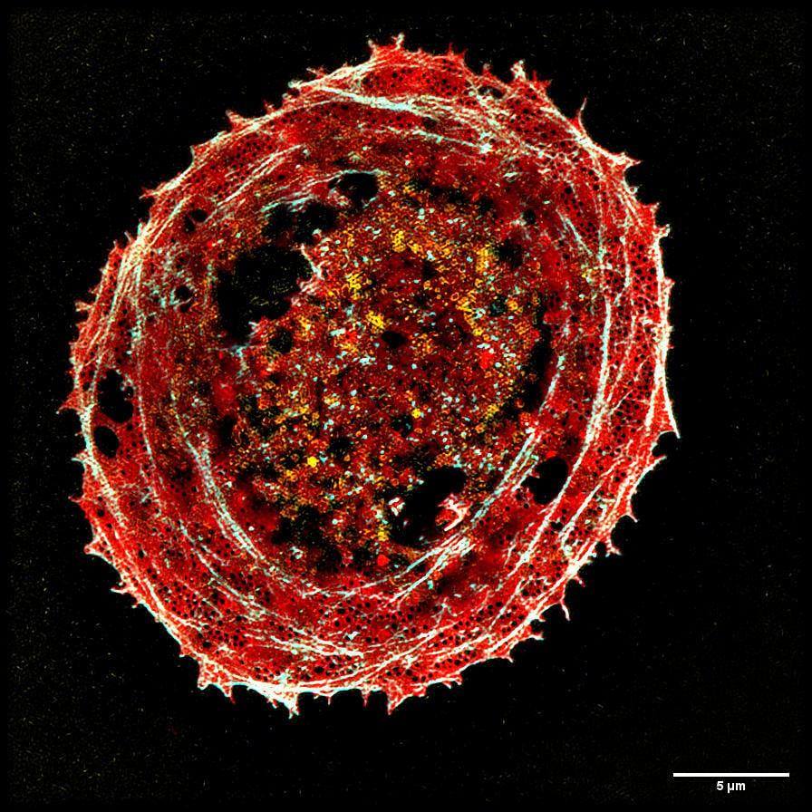
A new high-speed modulation 3DSIM system called "DMD-3DSIM" combines digital display with super-resolution imaging, enabling scientists to observe cellular structures in unprecedented detail. Developed by Professor Peng Xi's team at Peking University, this innovative setup utilizes a digital micromirror device and an electro-optic modulator to significantly improve both lateral and axial resolution, allowing for the capture of intricate details of subcellular structures and highly scattering plant cell ultrastructures. The hardware components and control mechanisms are openly available on GitHub, fostering collaboration and paving the way for the future of multidimensional imaging.
Researchers have developed a fluorescence microscope that uses structured illumination for fast super-resolution imaging over a wide field of view. This advanced microscope is designed to capture high-resolution images of multiple living cells simultaneously, enabling the analysis of how different drugs and their combinations affect the body. The microscope utilizes optical fiber delivery of excitation light to achieve high image quality and can perform multicolor and high-speed imaging. It has the potential to improve personalized healthcare and may find applications in clinical settings.