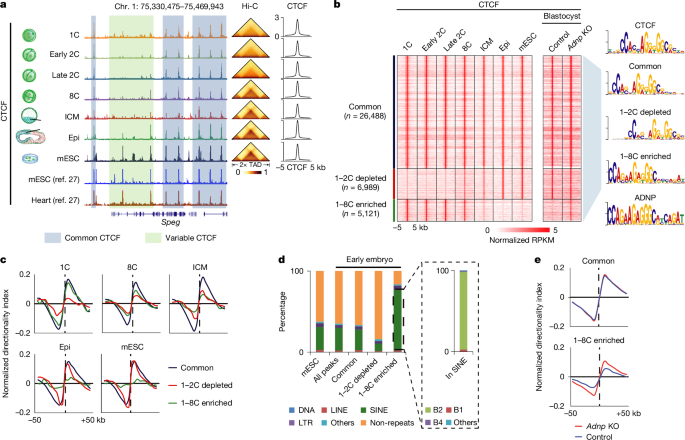
This study explores how chromatin structure influences hypertranscription during early embryonic development, utilizing extensive sequencing datasets and advanced genomic analysis tools to understand the interplay between chromatin architecture and gene expression in embryos.
Scientists have successfully created fertile mice with genetic material from two fathers through advanced gene editing techniques, marking a significant breakthrough in asexual reproduction research, though the process remains inefficient and raises ethical questions for future applications.
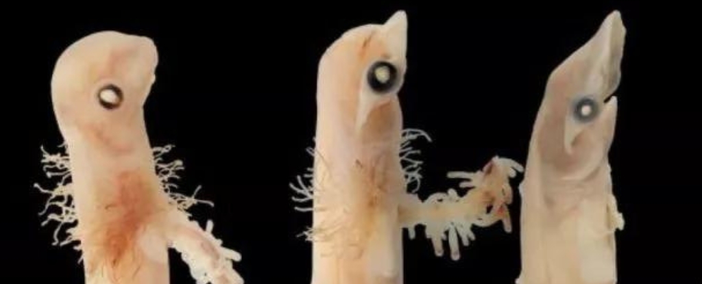
Scientists have gained rare access to the development of hammerhead shark embryos, shedding light on the mystery of their unique heads. Unlike most shark species, hammerheads gestate their pups in utero, with up to 16 embryos nourished by umbilical cords before live birth. By salvaging embryos from deceased adult female sharks, researchers were able to document the entire set of developmental stages, creating a visual growth chart and uncovering insights into the evolution of these peculiar sharks.
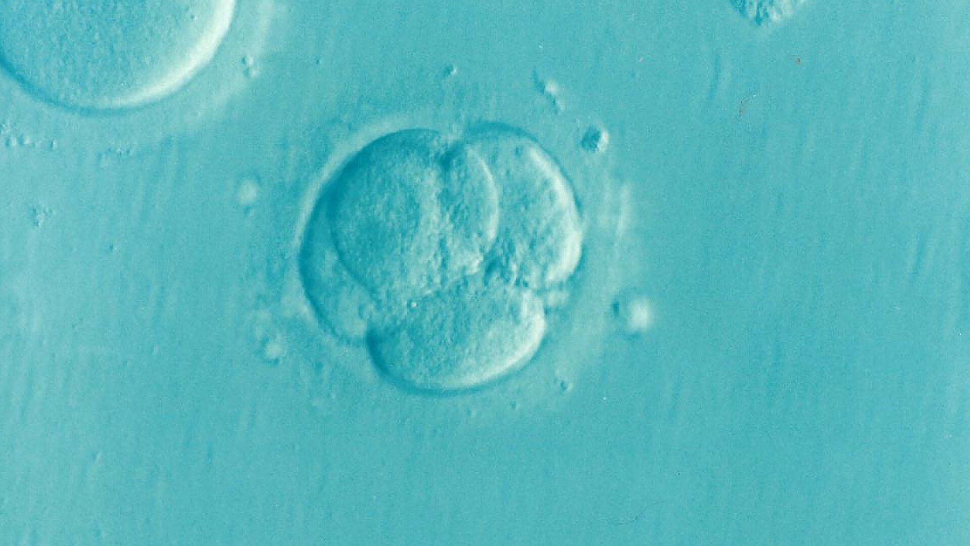
The Alabama Supreme Court ruling that frozen embryos can be considered "extrauterine children" under state law has significant implications for in vitro fertilization (IVF) procedures, potentially raising risks and costs for patients and medical practitioners. The ruling has led to at least three providers in Alabama pausing IVF treatments, and an Alabama lawmaker plans to introduce legislation to clarify IVF regulations. The ruling could impact the storage and use of frozen embryos, as well as the financial accessibility of IVF. Additionally, it may have broader implications for the recognition of embryos as persons in the context of the anti-abortion movement.
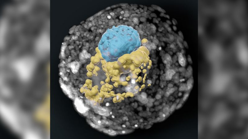
Researchers have discovered a link between a 500-million-year-old viral infection and the early stages of embryo development, shedding new light on the role of endogenous retroviruses in regulating pluripotency factors. The study, conducted on mouse embryos, identified a retroviral protein called MERVL-gag that influences the transition from totipotent to pluripotent cells, a crucial step in embryo specialization. This finding has implications for artificial embryo creation, regenerative medicine, and understanding fertility issues, highlighting the important functions of ancient retroviruses that have co-evolved with complex organisms over millions of years.
Researchers from the CNIO have discovered that a virus that infected animals hundreds of millions of years ago plays a crucial role in the development of embryos. The viral genetic material integrated into the genome of the first multi-cellular beings is still present in our DNA today and is essential for the transition from totipotency to pluripotency in embryos. This finding has implications for regenerative medicine and artificial embryo creation, as it opens up new possibilities for generating stable cell lines in the totipotency phases. The study sheds light on the symbiotic co-evolution of endogenous retroviruses with host cells to ensure the smooth progression of early embryonic development.
Dutch company Spaceborn United is conducting research on sex in space for the purpose of addressing the reproductive challenge of becoming a multi-planetary species. Their current focus is on conceiving a viable embryo in space, using a special disk to mix cells and cryogenically freezing the embryo for protection during reentry. While actual physical sex in space is still a ways off, the company plans to launch "mice cells" into space next year and a human embryo in five to six years. The ethical implications of implanting a space-fertilized embryo are a concern due to radiation and gravity differences. NASA is leaving such research to private firms like Spaceborn United, while space tourism may potentially beat them to the first sex in space.
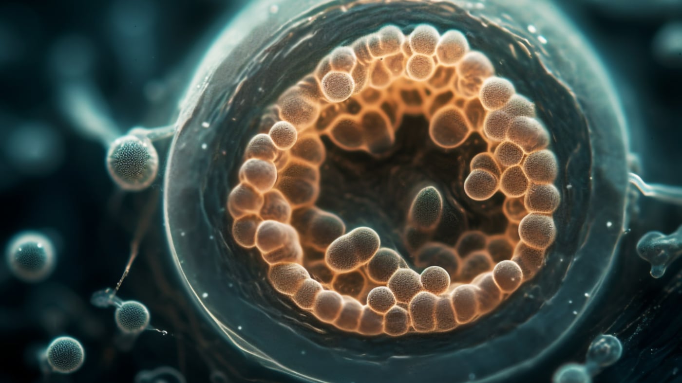
Recent breakthroughs in stem cell research have allowed scientists to create lab models of human embryos, providing insights into the crucial first month of development. These embryo-like structures, made from pluripotent stem cells, offer potential for understanding miscarriages, birth defects, and the effects of medications during pregnancy. While the models are not considered embryos and have limitations, they could serve as an ethical alternative to scarce human embryos for research purposes. However, concerns about ethical oversight and the potential for misuse remain, prompting the need for better regulation and guidelines in this emerging field.
Researchers have discovered that the OBOX gene family plays a crucial role in the activation of the zygote genome, initiating the development of an embryo. These genes guide the enzyme RNA polymerase II to transcribe the correct genes at the right time, allowing the embryo to develop successfully. The redundancy of these genes' functions ensures the critical transition from zygote to embryo occurs properly. This research provides new insights into the early stages of embryo development and may have implications for understanding embryonic stem cell reprogramming.
Rare genetic mutations that occur during the early stages of embryo development may increase the risk of developing schizophrenia later in life, according to researchers. These non-inherited mutations, known as somatic mutations, were found in genes NRXN1 and ABCB11. NRXN1 mutations were present in blood cells of individuals with schizophrenia and are believed to affect the brain's connections between nerve cells. ABCB11 mutations were found in dopamine-producing neurons and may impact the effectiveness of schizophrenia drugs. The findings could lead to new insights and potential treatments for the condition.
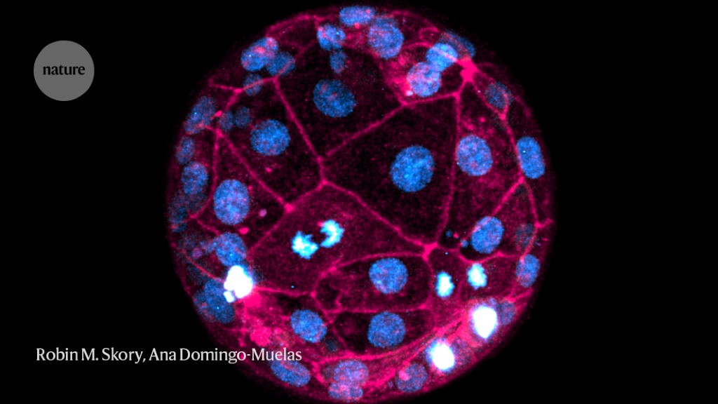
Researchers have developed a new imaging technique using fluorescent dyes and laser microscopes to capture the most detailed images of human embryos developing in real time. This non-invasive method allows for the study of crucial events in the early stages of development without genetically altering the embryos. The technique could have applications in non-invasive screening of embryos conceived through in vitro fertilization (IVF) and may provide insights into chromosomal abnormalities and variations in embryonic development between humans and mice. The researchers hope to further improve the imaging process and potentially use it in clinical settings to determine the viability of embryos before implantation.
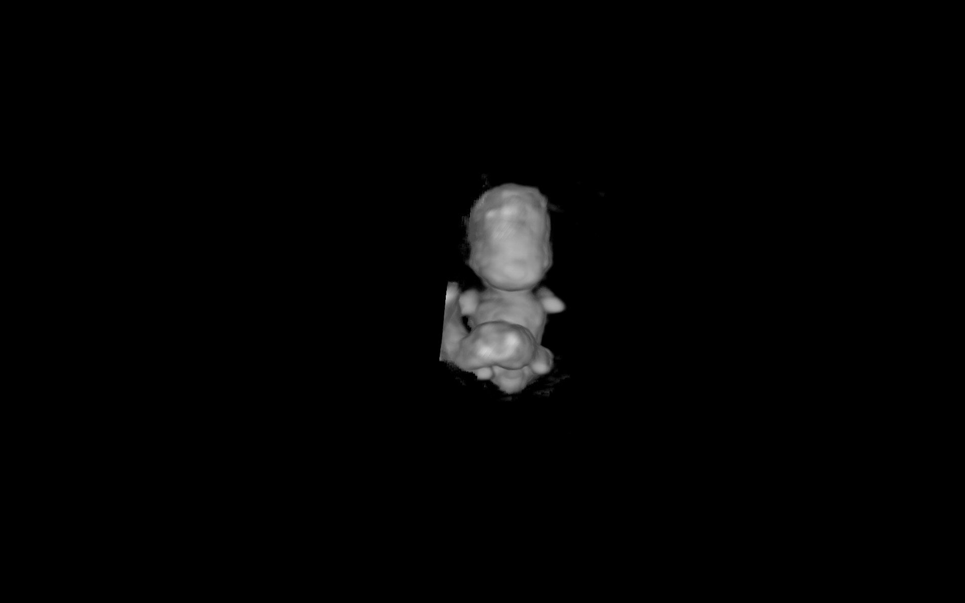
Embryos in pregnancies that end in miscarriage take longer to develop in the womb than those in pregnancies that result in live births, according to new research published in Human Reproduction. Researchers in The Netherlands used state-of-the-art imaging technology, including 3D ultrasound with high resolution transvaginal probes and virtual reality techniques, to create 3D holograms of the embryo and found that embryos in pregnancies that end in a miscarriage took four days longer to develop than babies that did not miscarry. The longer it takes for an embryo to develop, the more likely it is to miscarry. The ability to assess the shape and development of embryos could be used to estimate the likelihood of a pregnancy continuing to the delivery of a healthy baby.