Unprecedented Imaging Reveals Detailed Development of Model Human Embryos
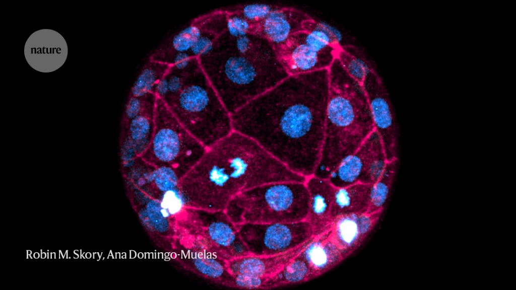
Researchers have developed a new imaging technique using fluorescent dyes and laser microscopes to capture the most detailed images of human embryos developing in real time. This non-invasive method allows for the study of crucial events in the early stages of development without genetically altering the embryos. The technique could have applications in non-invasive screening of embryos conceived through in vitro fertilization (IVF) and may provide insights into chromosomal abnormalities and variations in embryonic development between humans and mice. The researchers hope to further improve the imaging process and potentially use it in clinical settings to determine the viability of embryos before implantation.
Reading Insights
0
6
3 min
vs 4 min read
84%
650 → 103 words
Want the full story? Read the original article
Read on Nature.com