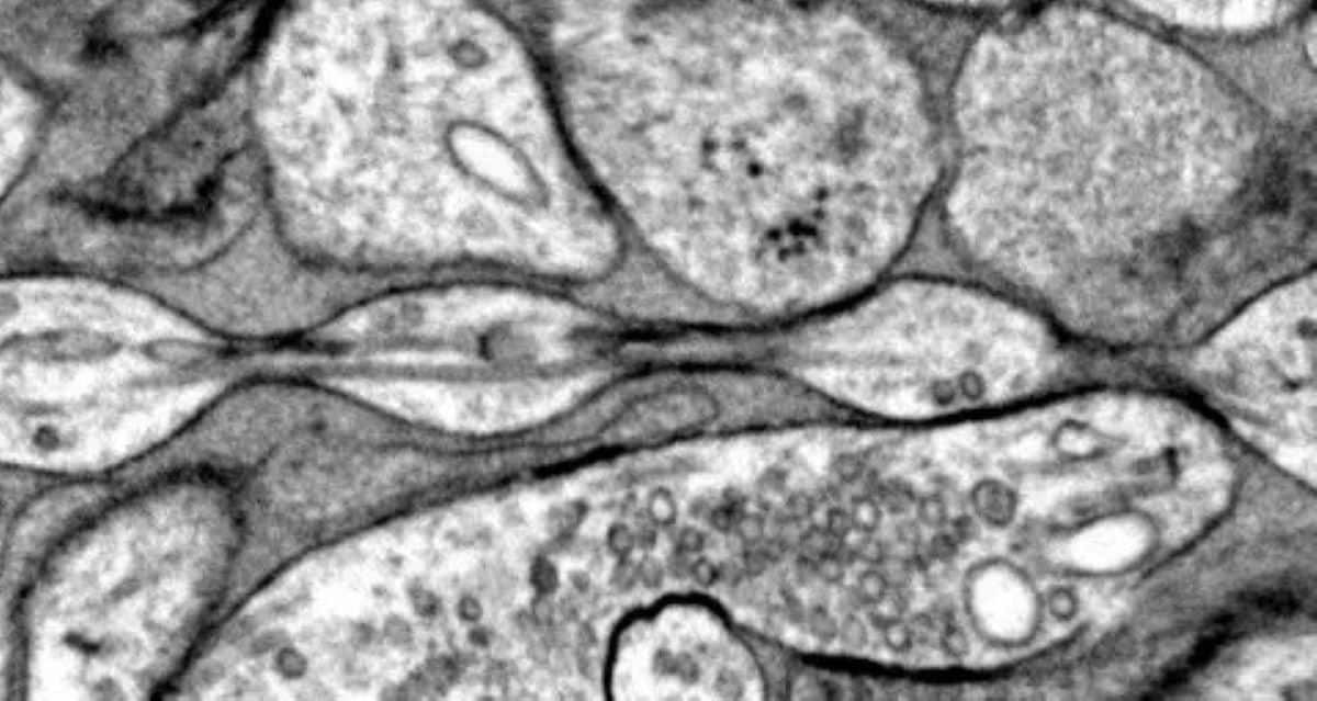
Researchers discovered that espresso coffee can effectively stain biological samples for electron microscopy, offering a safe, inexpensive, and non-toxic alternative to traditional heavy metal stains like uranyl acetate, with promising results in imaging zebrafish mitochondria, though further testing across different tissues is needed.
A study reveals that over 97% of electron microscope images are not published, representing a vast, untapped resource for AI and scientific discovery. Experts suggest systematic archiving and sharing of these images could enhance research and accelerate new findings.
A DIY project describes creating a calibration target for electron microscopes by depositing gold nanoparticles on silicon slides, enabling precise calibration and repair of the microscope's resolution capabilities.
A study using advanced electron microscopy reveals that axons, previously thought to be tube-like, have a "pearl-on-a-string" structure, termed "non-synaptic varicosities." This discovery, which may impact brain signaling and neurodegenerative disease research, shows that axon morphology is influenced by membrane mechanics, such as cholesterol levels. The findings challenge long-held beliefs about neuron structure and suggest potential revisions in biology textbooks.
Researchers in Germany have successfully sent single electrons along structured chiral paths, achieving chirality in electron matter waves without angular momentum. This work, which parallels earlier research with photons, could have significant applications in electron microscopy and the study of magnetic materials. However, some scientists are skeptical about the claim of chirality without angular momentum and the lack of citation of previous related work.
Physicists at the University of Konstanz have discovered a method to imprint chirality onto electrons using laser light, creating chiral coils of mass and charge. This breakthrough has significant implications for quantum optics, particle physics, and electron microscopy, potentially leading to new scientific explorations and technological advancements.
Researchers have discovered pristine extraterrestrial organic molecules, including amino acids and nucleobases, inside the Winchcombe meteorite, shedding light on the potential contributions to the development of life on Earth. This marks a significant advancement in understanding the solar system's formation and the role of carbonaceous meteorites in delivering organic compounds to the early Earth. The findings, published in Nature Communications, were made possible through advanced electron microscopy analysis, providing unprecedented resolution and efficiency in analyzing extraterrestrial organic matter.
Researchers at the University of Illinois Urbana-Champaign have demonstrated that record-breaking microscopic resolution can be achieved using electron ptychography on "conventional" transmission electron microscopes, breaking the trend of increasing microscope price with increasing resolution. This new technique, which uses computation to boost resolution, allows for deep sub-angstrom spatial resolution and rivals the highest ptychographic resolutions achieved with expensive aberration-corrected microscopes. The approach quadruples the resolution of conventional transmission electron microscopes and represents a significant paradigm shift in the field of electron microscopy.
Scientists from the University of Nottingham have successfully trapped individual krypton atoms inside carbon nanotubes, creating the world's first one-dimensional gas. Using advanced transmission electron microscopy, the team was able to directly image chains of noble gas atoms, a significant breakthrough in understanding individual atomic behavior. This innovation could lead to a better understanding of unusual states of matter and have wide-ranging effects on atomic-scale behavior, with potential applications in chemistry and physics.
Researchers have achieved a breakthrough by stabilizing and directly imaging small clusters of noble gas atoms, such as krypton and xenon, between two layers of graphene at room temperature. This discovery opens up new possibilities for research in condensed matter physics and potential applications in quantum information technology. The method involves trapping noble gas atoms between graphene layers, allowing for the observation of their behavior using scanning transmission electron microscopy. This development may lead to further studies on the properties of clusters with different noble gases and their potential applications in quantum technology.
Scientists have successfully integrated nonlinear optical phenomena, specifically "Kerr solitons," into a transmission electron microscope (TEM) using a photonic microresonator chip. These stable, localized pulses of light interacted with a beam of electrons, enabling ultrafast modulation of electron beams and demonstrating the potential for high repetition-rate ultrafast electron microscopy and particle accelerators on a small photonic chip. This breakthrough opens up new possibilities for probing nonlinear optical dynamics at the nanoscale and developing nonlinear photonic devices.
Researchers have developed a new technique for attosecond electron microscopy that allows for the observation of sub-cycle optical dynamics in materials. The technique involves using tilted electron pulses to image electromagnetic waveforms with unprecedented temporal resolution. This could lead to new insights into the behavior of plasmonic and chiral nanostructures, as well as the study of ultrafast electron dynamics in materials.
Researchers have used electron microscopy and atomic models to gain insight into the process by which the BAM complex mediates the assembly of outer membrane β-barrel proteins in bacteria. The study provides a structural basis for understanding the mechanism of protein assembly and could aid in the development of new antibiotics that target this process.
Researchers at University of Tokyo, JTS PRESTO, Ludwig Maximilians Universität and Kindai University have demonstrated the modulation of an electron source by applying laser light to a single fullerene molecule. The technique could enable the integration of ultrafast switches into a single fullerene molecule, paving the way for the development of better performing computers and microscopic imaging devices. The researchers also propose an integration scheme that would allow the integration of as many switches as desired without the need to increase the size of devices. The method could also be applied in the field of electron microscopy.
Researchers from Northwestern University and the University of Illinois have observed nanoparticles self-assembling into solid materials for the first time, offering valuable insights for the design of new materials, such as thin films for electronics. The study used a newly optimized form of liquid-phase transmission electron microscopy (TEM) to gain unprecedented insights into the self-assembly process. The researchers say this information will help engineers design new materials, specifically thin-film materials, which are often used to build flexible electronics, light-emitting diodes, transistors, and solar cells.