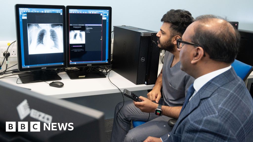
Leeds Teaching Hospitals NHS Trust has introduced AI 'co-pilot' software to accelerate diagnosis of conditions like lung cancer and infections through chest X-ray analysis, aiming to improve patient treatment times and safety, with the AI supporting doctors by identifying up to 85 findings quickly without replacing them.
Astronomers have used XMM-Newton, Chandra, and NuSTAR space telescopes to investigate the nature of a newly-discovered very-high-energy source, 2FHL J1745.1–3035, and found that it may be a powerful pulsar wind nebula (PWN). The source, located near the Galactic center, exhibits hard gamma-ray spectrum and is a TeV emitter. Analysis of X-ray properties suggests that it is compact with extended emission, and its broadband X-ray spectrum is best-fitted with a broken power law model. If confirmed, this would be one of the hardest PWNe ever detected in X-rays and the hardest in gamma-rays.
X-ray analysis of a tiny speck from the "Mona Lisa" has revealed new insights into Leonardo da Vinci's painting techniques. Scientists discovered a rare compound called plumbonacrite in the base layer of the painting, confirming that Leonardo likely used lead oxide powder to thicken and dry his paint. This finding provides scientific evidence for previously hypothesized techniques used by the artist. The research highlights Leonardo's experimental nature and the ongoing potential for further discoveries about his iconic masterpiece.
A Crusader sword discovered off the coast of Israel, believed to be from the 12th to 13th century, was found bent and covered in concretions. X-ray analysis revealed the sword's original outline and confirmed that it remained bent, suggesting it was damaged during the Crusades. The sword, known as the Newe-Yam sword, was likely used by a Crusader warrior who settled in the Holy Land after the First Crusade. Swords from this period are rare finds, and losing one during a naval battle would have been detrimental. The discovery provides insight into the battles fought between Crusaders and Muslims in the region.
X-ray analysis of a fossil of a marine reptile discovered in Edgeya, Svalbard, led to its categorisation which had not previously been done. The reptile most likely belongs to the ichthyosaur species Phalarodon atavus after X-ray imaging of the specimen revealed new information, including features of the skull and teeth. The study revealed that radiography works particularly well with the sulphur minerals that form fossils in the Norwegian archipelago. This approach might one day provide new information about ancient life.
X-ray analysis has led to the identification of a previously-unidentified marine reptile fossil discovered in Edgeøya, Svalbard, which lived around 240 million years ago. The study suggests that fossils from the Middle Triassic Botneheia Formation in Svalbard, Norway, are particularly suitable for radiographic imaging. The fossils have high X-ray contrast due to the presence of sulfate minerals, notably sulfate baryte, which could be linked to conditions created by ancient volcanic activity. This discovery not only demonstrates the utility of X-ray techniques for studying these fossils but also identifies conditions that can form fossils well-suited for these techniques, in Svalbard and potentially elsewhere.
X-ray analysis of a previously-unidentified marine reptile fossil from Svalbard, Norway has enabled its classification as the ichthyosaur species Phalarodon atavus. The Middle Triassic Botneheia Formation of Svalbard is particularly well-suited to radiographic imaging due to the high X-ray contrast provided by sulfate minerals, notably sulfate baryte. The formation of these minerals is little understood, but could be linked to conditions created by ancient volcanic activity. This study demonstrates the utility of X-ray techniques for studying fossils and identifies conditions that can form fossils well-suited for these techniques, in Svalbard and potentially elsewhere.
Scientists at The Australian National University and the Australian Museum Research Institute used micro-CT scans to examine the fossilized stomach remains of a small marine reptile, a plesiosaur nicknamed 'Eric,' to determine its diet. They found 17 previously undescribed fish vertebrae inside Eric's gut, confirming the plesiosaur's diet consisted mostly of fish. The study demonstrates the potential to use X-rays to reconstruct the diets of other extinct organisms that inhabited Earth hundreds of millions of years ago.