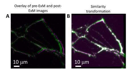
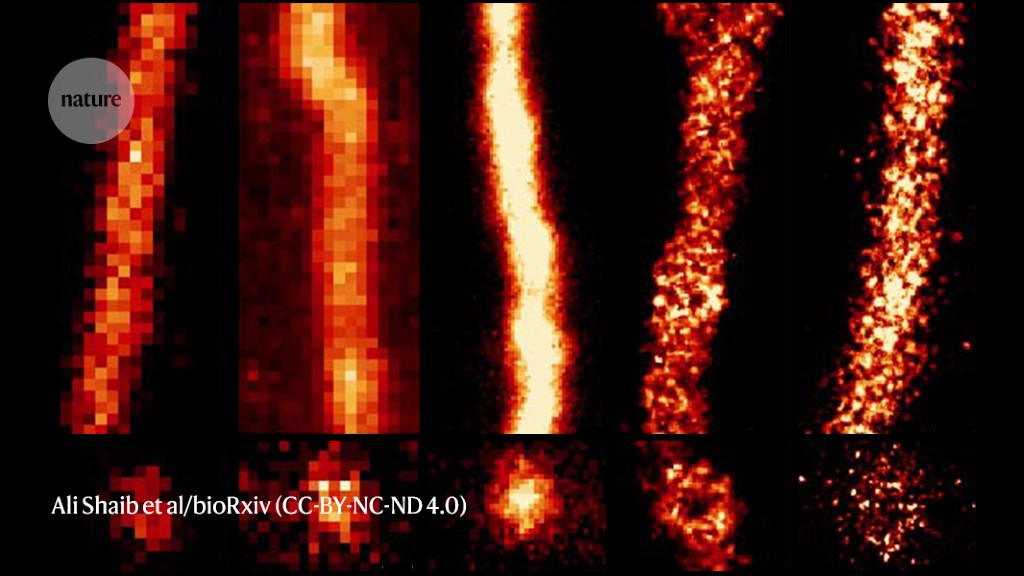
Expansion microscopy uses a diaper-inspired hydrogel to physically swell biological samples, enabling higher-resolution visualization of tiny cellular structures with standard microscopes. By improving dye penetration and preserving overall architecture, it democratizes microscopy and reveals detailed cytoskeletal diversity across species.
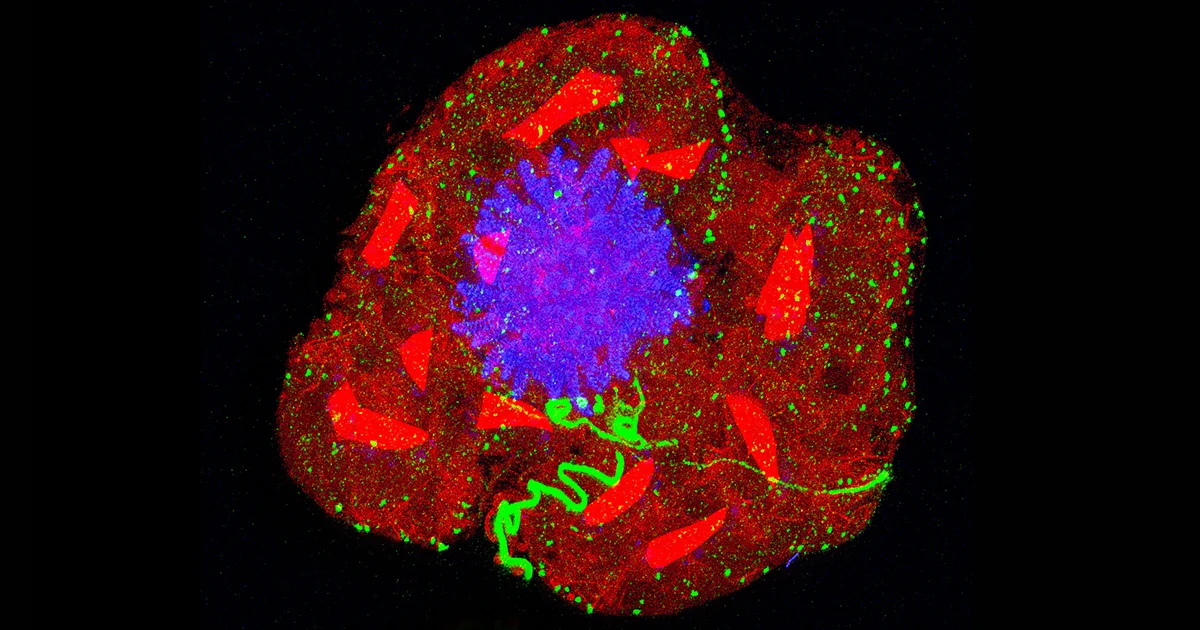
Researchers at MIT and Brigham and Women’s Hospital/Harvard Medical School have developed a novel microscopy technique that allows for high-resolution imaging of human brain tissue, revealing previously unseen cells and structures. The method, based on expansion microscopy, has potential applications in diagnosing tumors, providing more accurate prognoses, and guiding treatment choices. By labeling up to 16 different molecules per tissue sample, the researchers were able to analyze healthy brain tissue and samples from patients with gliomas, uncovering unexpected levels of aggressive tumor cells in low-grade gliomas. The technique could serve as a diagnostic tool for neuro-oncology and neuropathology, offering insights into neurological diseases at the nanoscale and potentially leading to improved patient outcomes.
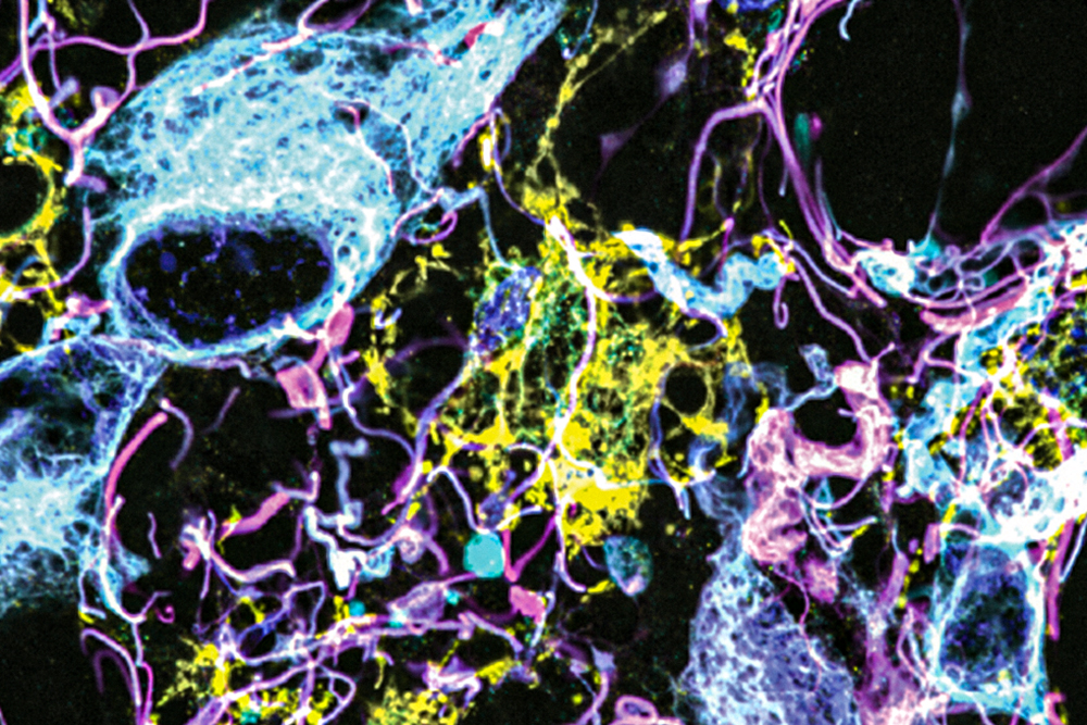
Researchers at Washington University in St. Louis have developed a new technique called plasmon-enhanced expansion microscopy (p-ExM) that improves the resolution of imaging very small objects like neurons. By using ultrabright fluorescent markers called plasmonic-fluors (PFs), the team was able to overcome the issue of signal loss during the expansion process. The PFs, constructed from gold and silver nanoparticles, significantly increased the brightness of the fluorophores and allowed for clearer imaging of neural networks. The technique has the potential to aid in mapping connections between neurons and other high-resolution imaging applications.
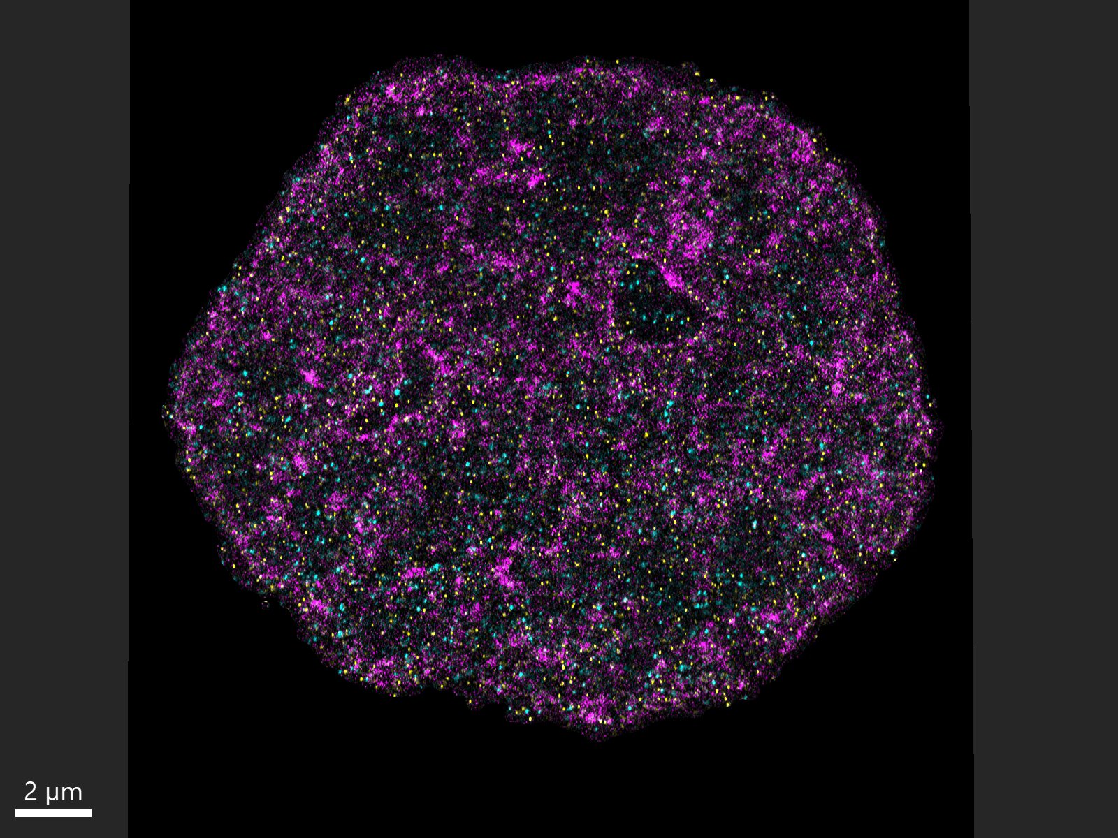
Scientists at Yale University have developed a new microscopy technique called chromatin expansion microscopy (ChromExM) that allows them to visualize previously unseen molecular processes within genetic material. By expanding the physical volume of nuclei in zebrafish embryonic cells, the researchers were able to drastically improve image resolution and observe how individual molecules shape gene expression during embryonic development. This breakthrough technique, which provides valuable insights into gene regulation, could lead to a better understanding of fundamental processes in the nucleus and have implications for various fields, from embryology to cancer research.
Researchers at the University Medical Center Göttingen in Germany have developed a new microscopy technique called ONE microscopy that can achieve resolutions below 1 nm, revealing the shape of individual proteins. The technique combines the physics-beating super-resolution methods and expansion microscopy to offer a level of detail that eclipses even that of multi-million-dollar ‘super-resolution’ microscopes. The ONE microscopy technique is straightforward to apply and works with now-antiquated fluorescent microscopes from the 1990s, making it accessible to researchers in low and middle-income countries.