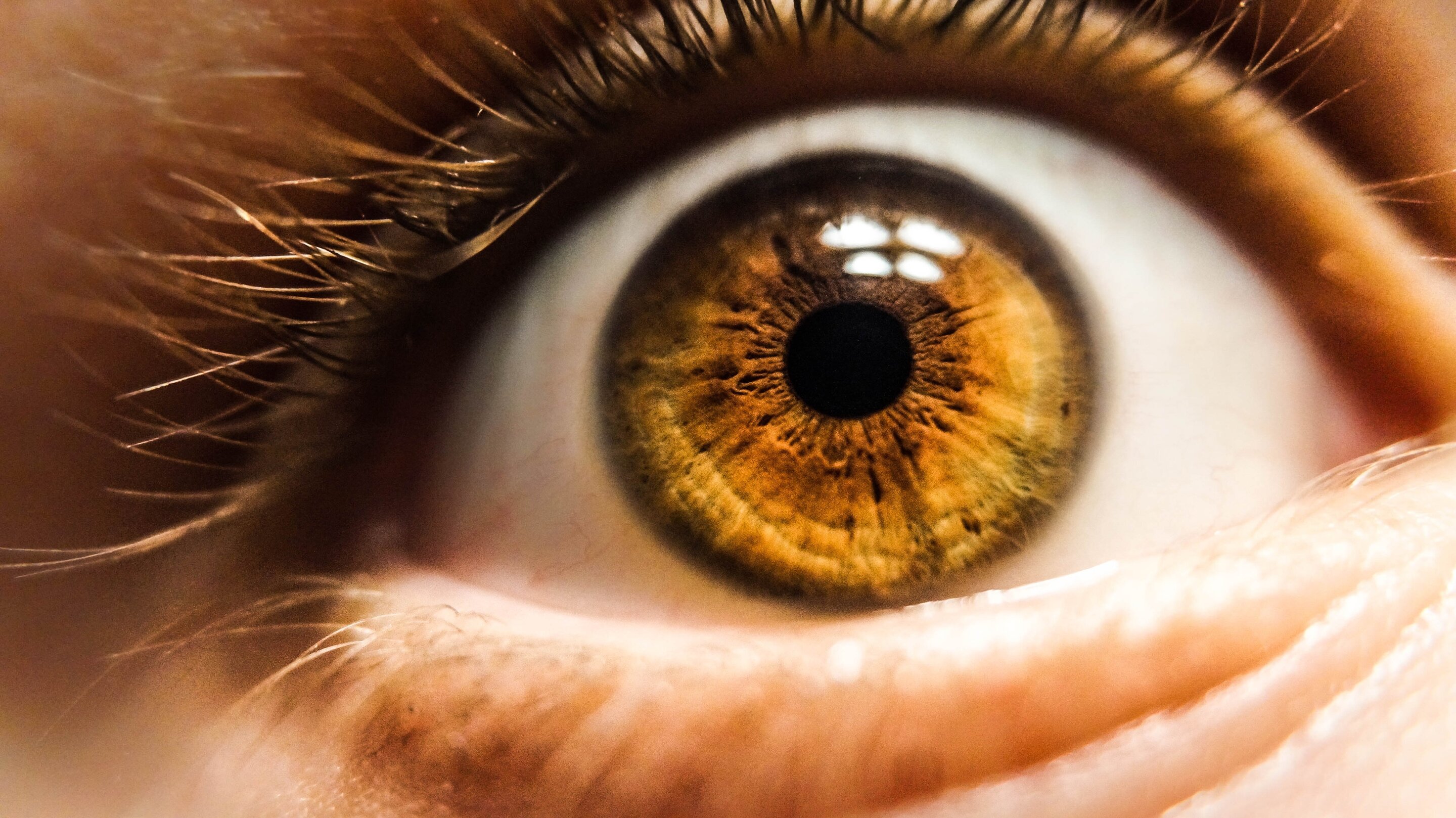
Oculomics is an emerging field that uses AI and high-resolution retinal images to detect systemic diseases like heart disease, diabetes, and neurological disorders early, by analyzing subtle eye features that reflect overall health, potentially transforming preventive medicine.
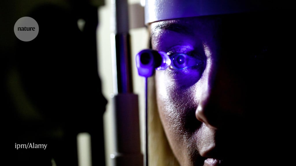
Routine eye exams, especially retinal imaging like OCT scans, can reveal early signs of heart disease by detecting vascular changes in the retina, enabling earlier intervention and prevention of serious cardiovascular events.
Researchers at the National Institutes of Health have utilized artificial intelligence (AI) to significantly improve the speed and quality of retinal cell imaging, making it 100 times faster and enhancing image contrast 3.5-fold. By integrating AI with adaptive optics optical coherence tomography (AO-OCT), they have developed a deep learning algorithm called P-GAN, which successfully de-speckles images of the retinal pigment epithelium (RPE) cells, reducing imaging acquisition and processing time by about 100-fold. This advancement provides a better tool for evaluating age-related macular degeneration (AMD) and other retinal diseases, potentially revolutionizing clinical imaging and research in the field.
Researchers at the National Institutes of Health have utilized artificial intelligence (AI) to significantly improve the speed and quality of retinal imaging, making it 100 times faster and enhancing image contrast 3.5-fold. By integrating AI with adaptive optics optical coherence tomography (OCT), the new method, called parallel discriminator generative adverbial network (P-GAN), successfully de-speckles retinal pigment epithelium (RPE) images, providing a better tool for evaluating age-related macular degeneration and other retinal diseases. This advancement is expected to make AO imaging more accessible for routine clinical applications and studies aimed at understanding blinding retinal diseases.
Scientists have developed an AI tool called RETFound that can diagnose and predict the risk of various health conditions, including ocular diseases, heart failure, and Parkinson's disease, based on retinal images. The tool was developed using self-supervised learning, similar to large-language models like ChatGPT. By training on unlabelled retinal images, RETFound can learn to predict missing portions of images and classify them for specific conditions. The model has shown promising results in detecting ocular diseases and outperforms other AI models in predicting the risk of systemic diseases. The researchers have made the model publicly available for adaptation and training in different medical settings, but caution is needed to ensure ethical and safe usage.
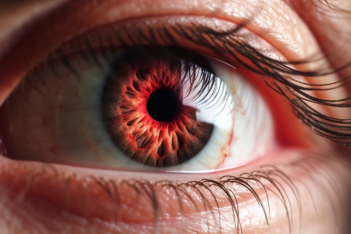
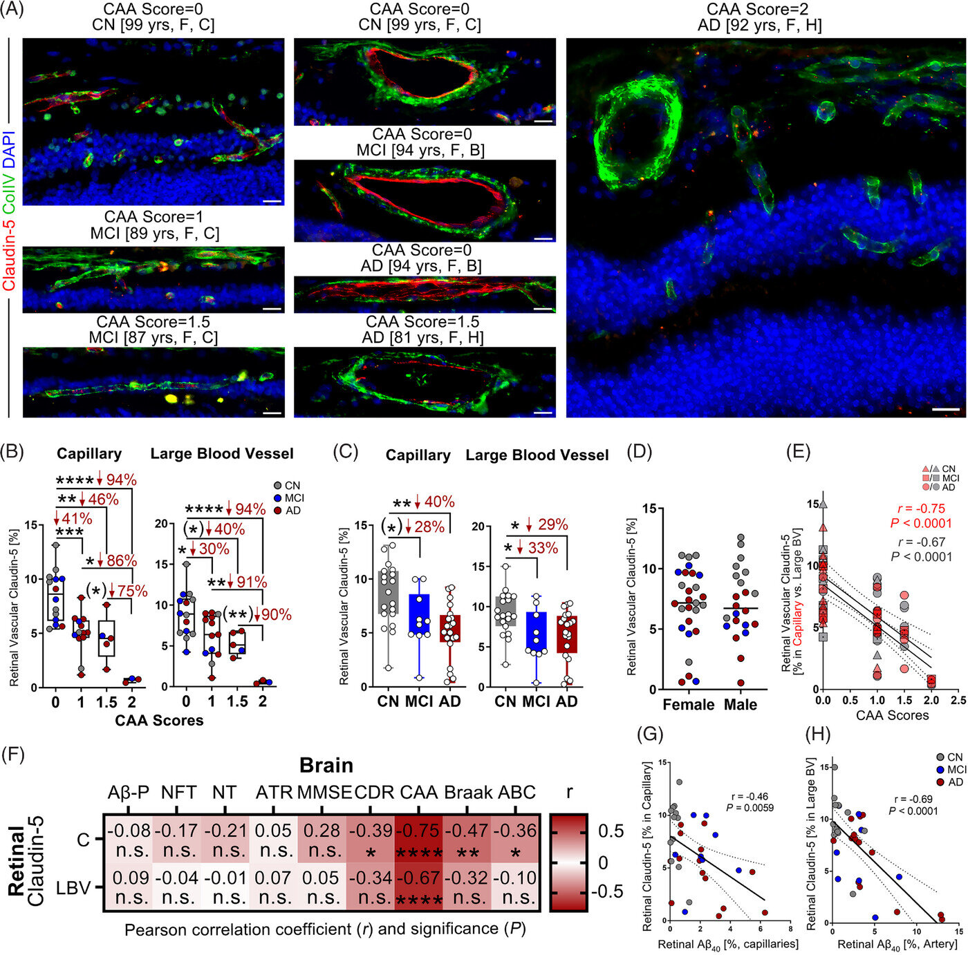
Abnormalities in eye blood vessels, specifically a disruption in the blood-retinal barrier, could provide early diagnosis of Alzheimer’s disease and tracking of its progression. The damage to the blood-retinal barrier was strongly linked with cerebral amyloid angiopathy (CAA), a condition associated with Alzheimer’s. Advanced retinal imaging, currently under development, could noninvasively monitor these changes in eye blood vessels to detect Alzheimer’s in living patients.
Blood vessel abnormalities in the eye are a major factor in the progression of Alzheimer's disease, according to research from Cedars-Sinai investigators. These changes correspond to changes in the brain, offering a new possibility for early diagnosis. Investigators compared blood vessels in retinas collected from 24 human donors with Alzheimer's disease, 10 donors with mild cognitive impairment and 27 with normal cognition. In patients with Alzheimer's disease and mild cognitive impairment, they found one of the earliest signs of Alzheimer's disease to date: disruption of the blood-retinal barrier, where tightly joined cells prevent harmful substances from entering the retinal tissue.