"Revolutionary Imaging Method Uncovers Hidden Brain Structures"
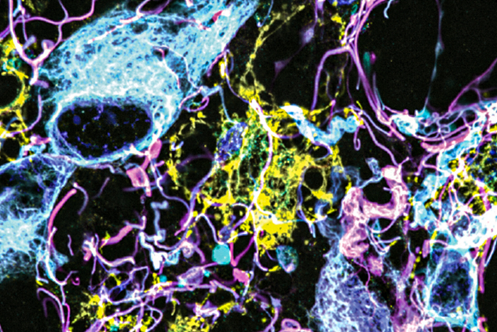
Researchers at MIT and Brigham and Women’s Hospital/Harvard Medical School have developed a novel microscopy technique that allows for high-resolution imaging of human brain tissue, revealing previously unseen cells and structures. The method, based on expansion microscopy, has potential applications in diagnosing tumors, providing more accurate prognoses, and guiding treatment choices. By labeling up to 16 different molecules per tissue sample, the researchers were able to analyze healthy brain tissue and samples from patients with gliomas, uncovering unexpected levels of aggressive tumor cells in low-grade gliomas. The technique could serve as a diagnostic tool for neuro-oncology and neuropathology, offering insights into neurological diseases at the nanoscale and potentially leading to improved patient outcomes.
Reading Insights
0
0
6 min
vs 7 min read
91%
1,247 → 113 words
Want the full story? Read the original article
Read on MIT News