MIT researchers have developed a noninvasive, Raman spectroscopy-based device that can measure blood glucose levels without finger pricks, showing promise as a comfortable alternative for diabetes management, with ongoing efforts to miniaturize and adapt it for wearable use.
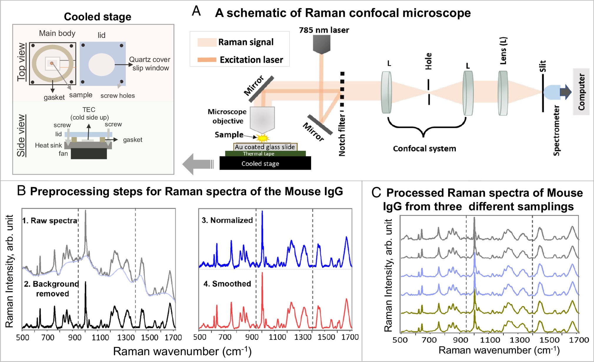
Researchers from Texas A&M University and TEES have developed a new technique called thermostable-Raman-interaction-profiling (TRIP) that overcomes the limitations of Raman spectroscopy in damaging live proteins during optical measurements. TRIP allows low-concentration, low-dose screenings for protein-to-ligand interactions in physiologically relevant conditions, enabling label-free, highly reproducible measurements. This breakthrough has potential applications in rapid and cost-effective drug, vaccine, and virus testing, as well as DNA analysis. The TRIP technique offers real-time detection of protein-ligand interactions, shortening the timeline for testing and providing accurate results. Additionally, the TRIP method requires smaller sample sizes and lower protein concentrations, making it a more cost-effective process.
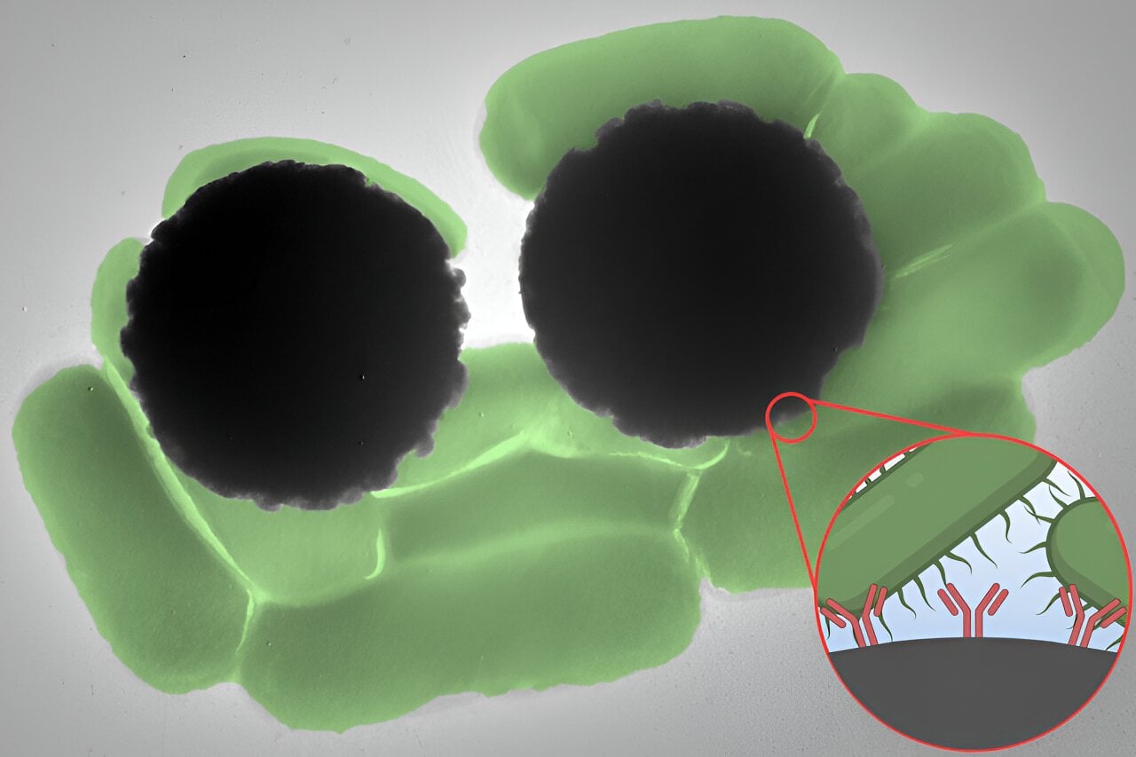
Researchers at MIT have discovered that Dynabeads, antibody-coated superparamagnetic beads, have a strong Raman signature that can be used to quickly detect pathogens in various diagnostic tests. By using Raman spectroscopy, the researchers were able to confirm the presence of Dynabead-bound pathogens within less than an hour, providing a rapid and reliable method for detecting contaminants such as Salmonella. The team is now working on developing a portable device for detecting bacterial pathogens, which could have significant applications in healthcare and resource-limited environments.
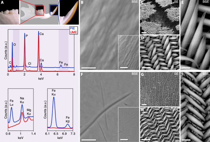
High-resolution Raman spectroscopy was used to analyze the compositional differences between pigmented incisor enamel (PIE) and unpigmented molar enamel (UME) in rats. The study found that PIE had higher levels of iron, higher mineral crystallinity, and higher acid phosphate and hydroxyl levels compared to UME. The enamel prisms in PIE were also wider and longer than those in UME. These findings provide new insights into enamel biomineralization and highlight the differences in ameloblast function between incisors and molars in rodents.
Researchers at Texas A&M University have developed a new technique called thermostable-Raman-interaction-profiling (TRIP) that overcomes the limitations of Raman spectroscopy in studying live proteins. By cooling the surface or substrate, the proteins remain intact during optical measurements, leading to highly reproducible results. This breakthrough has significant implications for drug and vaccine testing, as well as clinical diagnostics, by shortening the timeline and improving accuracy. The TRIP technique also requires smaller sample sizes and lower protein concentrations, making it a more cost-effective process. Future applications may include DNA analysis and other biological molecules.