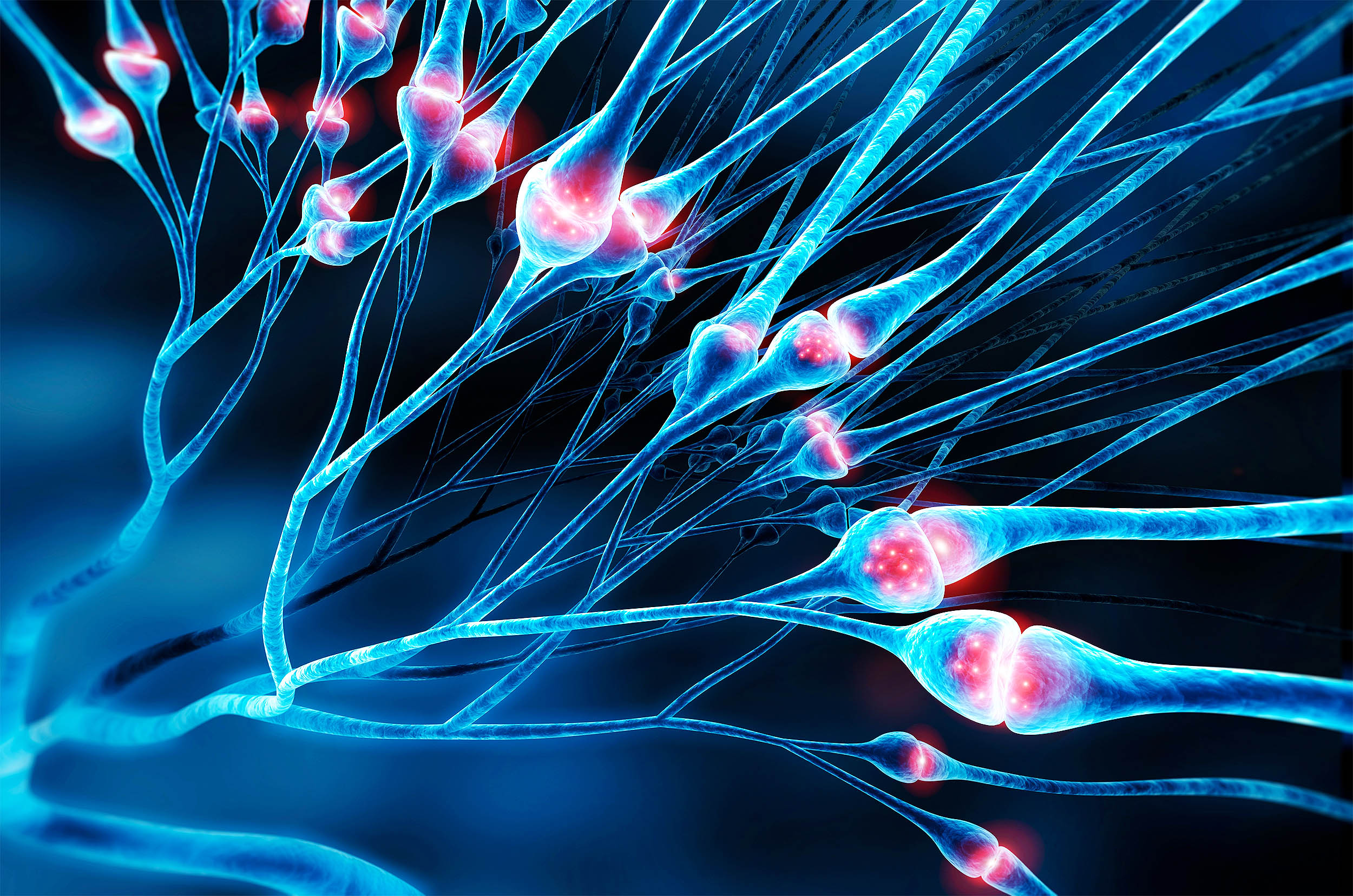
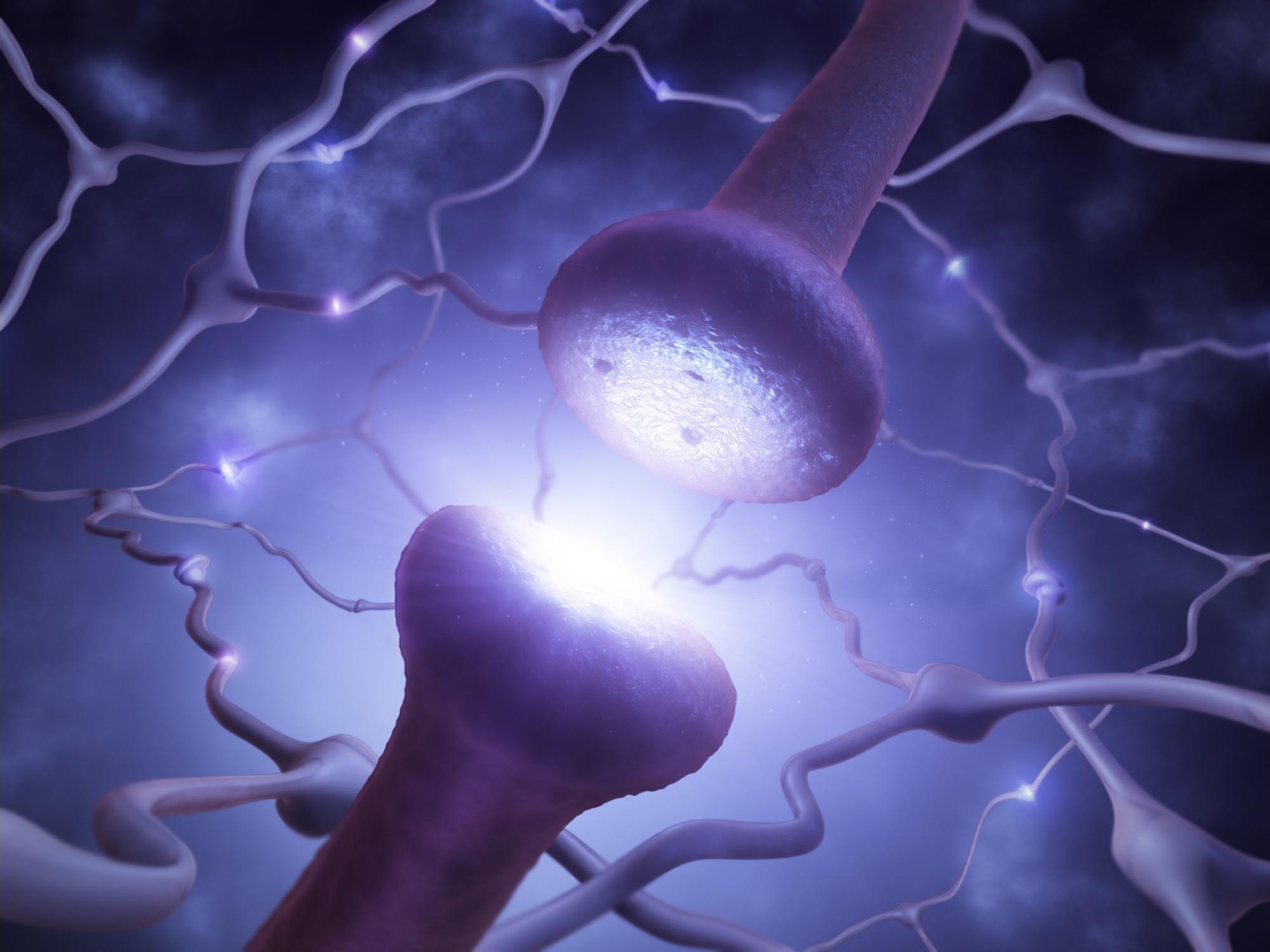
Researchers have discovered that the human retina actively synchronizes visual signals before they reach the brain by adjusting nerve fiber diameters and conduction speeds, ensuring a unified and precise visual experience. This process begins in the retina itself, challenging previous assumptions that synchronization occurs only in the brain.
New research using advanced imaging techniques reveals that axons, previously thought to be smooth tubes, are often shaped like strings of tiny bubbles or pearls, challenging a century-old understanding and suggesting a more dynamic and flexible structure that influences neural signaling.
Johns Hopkins researchers have discovered that axons in brain cells resemble a string of pearls rather than smooth tubes, challenging a century-old assumption about neuron structure. This finding, published in Nature Neuroscience, suggests that axon shape significantly impacts brain signaling and could influence our understanding of neurodegenerative diseases. The study used advanced imaging techniques and mathematical modeling to explore how membrane mechanics affect axon morphology and function, potentially necessitating revisions in biology textbooks.
MIT scientists found that the protein perlecan is vital for maintaining the structural integrity of neuronal axons. Without it, axons can break, leading to the death of synapses. Perlecan is part of the extracellular matrix that surrounds cells and helps them develop in a supportive, yet non-rigid environment. Humans need at least some perlecan to survive after birth. Mutations that reduce, but don’t eliminate, perlecan can cause Schwartz-Jampel syndrome, in which patients experience neuromuscular problems and skeletal abnormalities.