Unveiling the Structure of the Human Cardiac Myosin Filament
TL;DR Summary
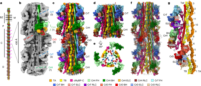
Researchers have used cryo-electron microscopy (cryo-EM) to determine the structure of the human cardiac myosin filament, a key component of muscle contraction in the heart. The study provides insights into the organization and arrangement of proteins within the filament, including myosin heads, tails, titins, and cMyBP-C. The structural data has been deposited in public databases, allowing other scientists to access and analyze the findings. This research contributes to our understanding of the molecular mechanisms underlying heart function and may have implications for the development of treatments for cardiac diseases.
Topics:health#cardiac-myosin-filament#cryo-em#human-heart#protein-structure#science-and-technology#structural-biology
- Cryo-EM structure of the human cardiac myosin filament Nature.com
- Scientists shoot first true-to-life 3D image of the thick filament of mammalian heart muscle Phys.org
- Structure of the native myosin filament in the relaxed cardiac sarcomere Nature.com
- Getting to the heart of thick-filament structure Nature.com
- View Full Coverage on Google News
Reading Insights
Total Reads
0
Unique Readers
16
Time Saved
37 min
vs 38 min read
Condensed
99%
7,539 → 89 words
Want the full story? Read the original article
Read on Nature.com