Unveiling the Neural Correlates of Rumination through Brain Imaging
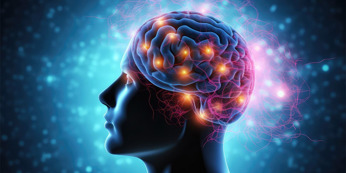
Researchers have used resting-state functional magnetic resonance imaging (rsfMRI) to identify the dorsal medial prefrontal cortex (dmPFC) as a key region involved in rumination, a mental process characterized by persistent negative self-reflective thoughts. By analyzing brain connectivity, the study found that the dmPFC interacts with other brain regions, such as the left inferior frontal gyrus (IFG) and right temporoparietal junction (TPJ), indicating language-based rumination and continuous evaluation of social scenarios. The study provides insights into the neural underpinnings of rumination and its potential links to depression and anxiety, offering a promising step towards understanding and treating these mental disorders.
Reading Insights
0
1
2 min
vs 3 min read
81%
528 → 99 words
Want the full story? Read the original article
Read on PsyPost