Unveiling the Intricacies of Cardiac Myosin Filament Structure
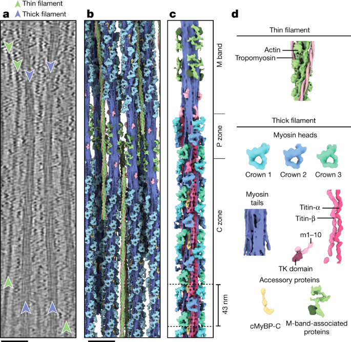
Researchers have determined the structure of the native myosin filament in the relaxed cardiac sarcomere, the basic contractile unit of muscle. The study reveals the arrangement and interactions of key proteins, including myosin, titin, and MyBP-C, within the thick filament. The myosin heads are organized in a quasihelical array with three-fold rotational symmetry, and the myosin tails form a coiled-coil structure at the center of the filament. MyBP-C acts as a mechanical sensor and links the thick and thin filaments, while titin acts as a molecular spring and ruler for myosin assembly. The findings provide insights into the molecular mechanics of muscle contraction and could have implications for understanding and treating cardiac diseases.