Unveiling the Intricate Details of the Human Retina through High-Resolution Imaging.
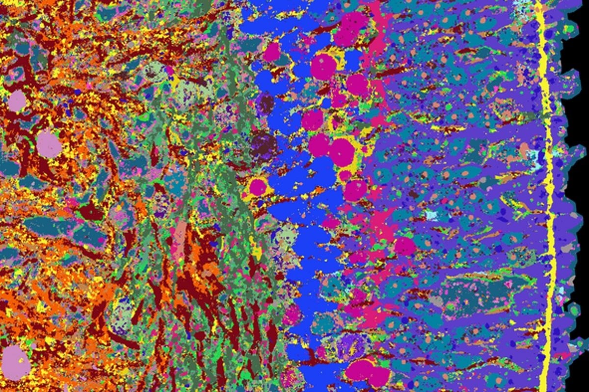
Researchers developed a new imaging technique called iterative indirect immunofluorescence imaging (4i) to visualize several dozen proteins in a thin tissue section at high resolution using fluorescence microscopy. The technique enabled researchers to map the development of human retinal organoids at high temporal and spatial resolution, providing insights into how healthy tissue forms and developing a time series that describes the entire 39-week development of retinal organoids. The researchers aim to apply this approach to other tissue types, such as the human brain and various tumor tissues, creating an atlas that provides information on the development of human organoids and tissues.